Copyright
©The Author(s) 2024.
World J Clin Oncol. Aug 24, 2024; 15(8): 1102-1109
Published online Aug 24, 2024. doi: 10.5306/wjco.v15.i8.1102
Published online Aug 24, 2024. doi: 10.5306/wjco.v15.i8.1102
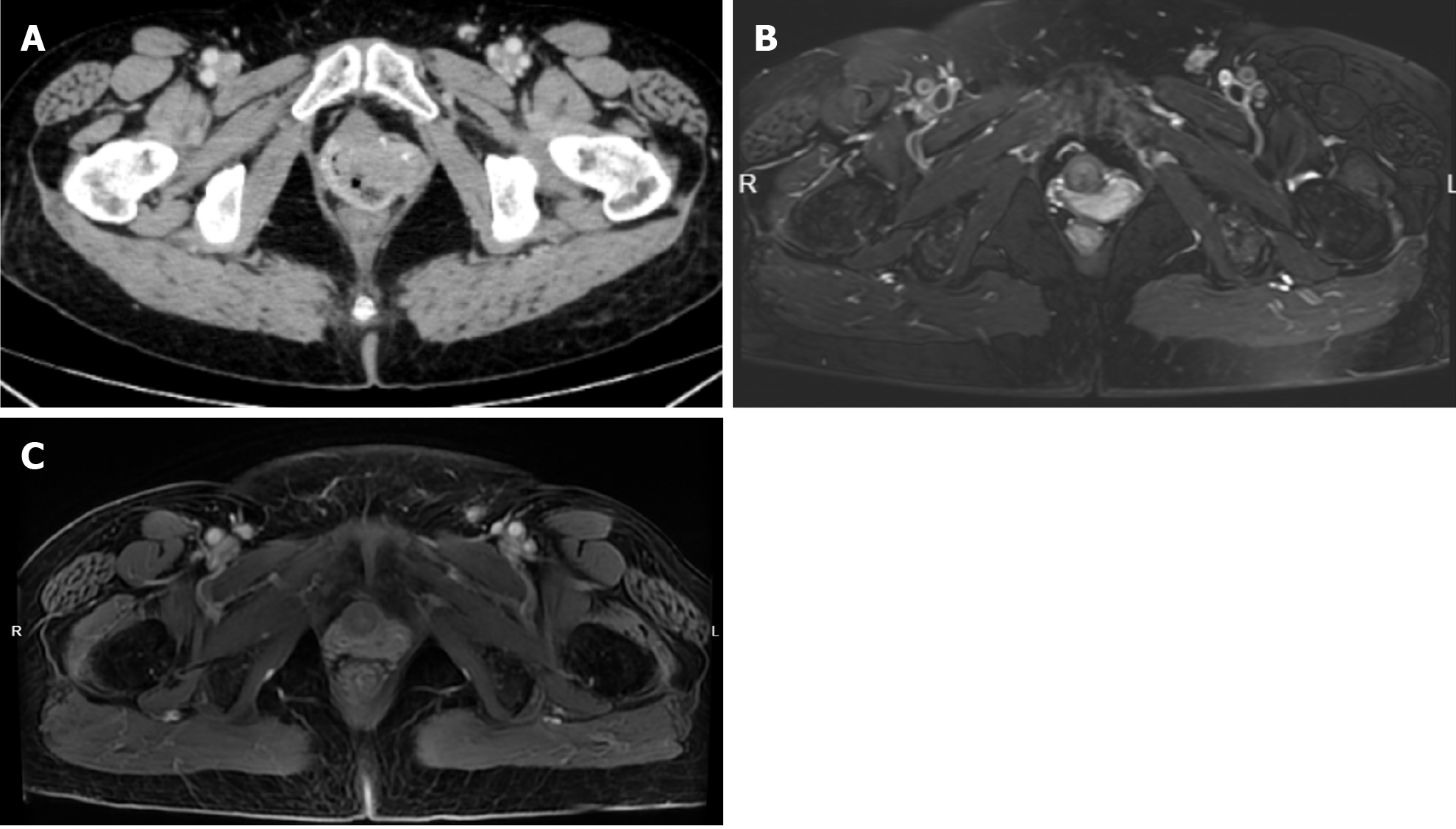
Figure 3 Morphological assessment.
A: Pelvic enhanced computed tomography before treatment shows occupation of the left and anterior vaginal walls, maximum cross-section range 40 mm × 30 mm; B: Enhanced magnetic resonance image after 3 cycles of chemotherapy shows thickening of the left anterior vaginal wall; C: Enhanced magnetic resonance image after external irradiation.
- Citation: Saijilafu, Gu YJ, Huang AW, Xu CF, Qian LW. Individualized vaginal applicator for stage IIb primary vaginal adenocarcinoma: A case report. World J Clin Oncol 2024; 15(8): 1102-1109
- URL: https://www.wjgnet.com/2218-4333/full/v15/i8/1102.htm
- DOI: https://dx.doi.org/10.5306/wjco.v15.i8.1102


