Copyright
©The Author(s) 2024.
World J Clin Oncol. Jun 24, 2024; 15(6): 755-764
Published online Jun 24, 2024. doi: 10.5306/wjco.v15.i6.755
Published online Jun 24, 2024. doi: 10.5306/wjco.v15.i6.755
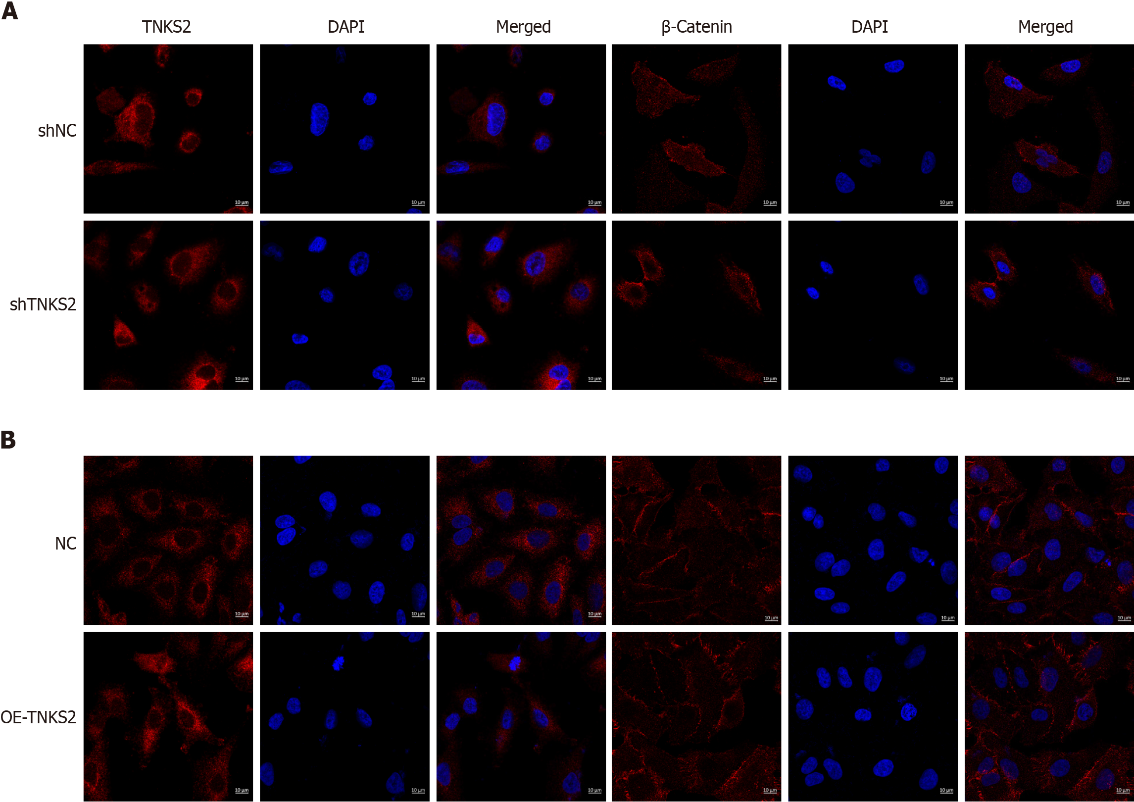
Figure 3 Immunofluorescence images obtained using confocal laser scanning microscopy.
A: Tankyrase 2 (TNKS2) and β-catenin expression in shTNKS2-transfected H647 cells; B: TNKS2-overexpressing A549 cells (magnification, × 200). Nuclei were stained blue; the target protein was stained red.
- Citation: Wang Y, Zhang YJ. Tankyrase 2 promotes lung cancer cell malignancy. World J Clin Oncol 2024; 15(6): 755-764
- URL: https://www.wjgnet.com/2218-4333/full/v15/i6/755.htm
- DOI: https://dx.doi.org/10.5306/wjco.v15.i6.755


