Copyright
©The Author(s) 2024.
World J Clin Oncol. May 24, 2024; 15(5): 653-663
Published online May 24, 2024. doi: 10.5306/wjco.v15.i5.653
Published online May 24, 2024. doi: 10.5306/wjco.v15.i5.653
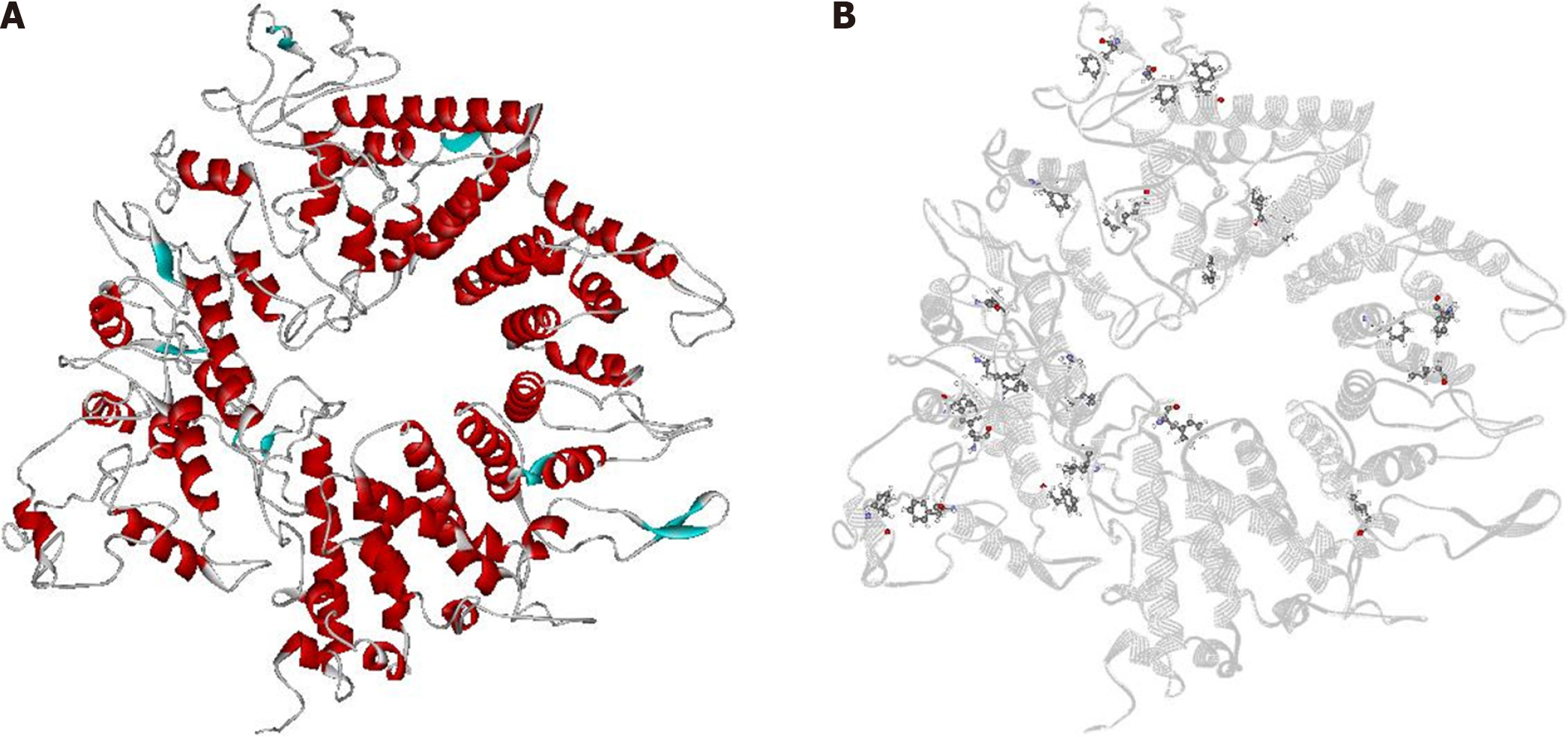
Figure 1 Three-dimensional structure model of Helicobacter pylori's CagA and predicted binding sites in silico.
A: CagA structure model. Blue: β-sheets; red: α-helices; gray: loops; B: CagA structure model in gray flat ribbon with predicted binding sites (residues) highlighted in ball and stick format.
- Citation: Vieira RV, Peiter GC, de Melo FF, Zarpelon-Schutz AC, Teixeira KN. In silico prospective analysis of the medicinal plants activity on the CagA oncoprotein from Helicobacter pylori. World J Clin Oncol 2024; 15(5): 653-663
- URL: https://www.wjgnet.com/2218-4333/full/v15/i5/653.htm
- DOI: https://dx.doi.org/10.5306/wjco.v15.i5.653


