Copyright
©The Author(s) 2024.
World J Clin Oncol. Nov 24, 2024; 15(11): 1435-1443
Published online Nov 24, 2024. doi: 10.5306/wjco.v15.i11.1435
Published online Nov 24, 2024. doi: 10.5306/wjco.v15.i11.1435
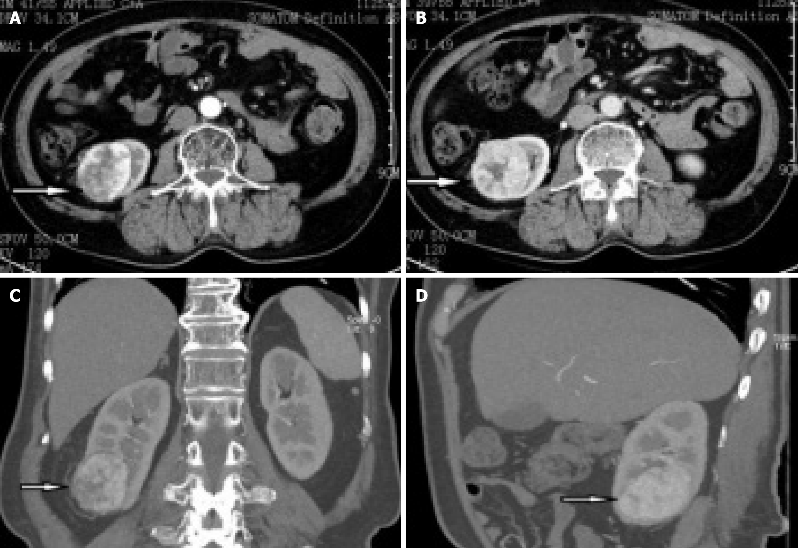
Figure 1 Abdominal contrast-enhanced computed tomography scans revealed that a heterogeneous mass was seen in the lower pole of the right kidney, with the size of about 53 mm × 47 mm.
A: Arterial phase; B: Venous phase; C: Coronal section; D: Median sagittal section.
- Citation: Tang JE, Wang RJ, Fang ZH, Zhu PY, Yao JX, Yang H. Treatment of fat-poor renal angiomyolipoma with ectopic blood supply by fluorescent laparoscopy: A case report and review of literature. World J Clin Oncol 2024; 15(11): 1435-1443
- URL: https://www.wjgnet.com/2218-4333/full/v15/i11/1435.htm
- DOI: https://dx.doi.org/10.5306/wjco.v15.i11.1435


