Copyright
©The Author(s) 2024.
World J Clin Oncol. Oct 24, 2024; 15(10): 1366-1375
Published online Oct 24, 2024. doi: 10.5306/wjco.v15.i10.1366
Published online Oct 24, 2024. doi: 10.5306/wjco.v15.i10.1366
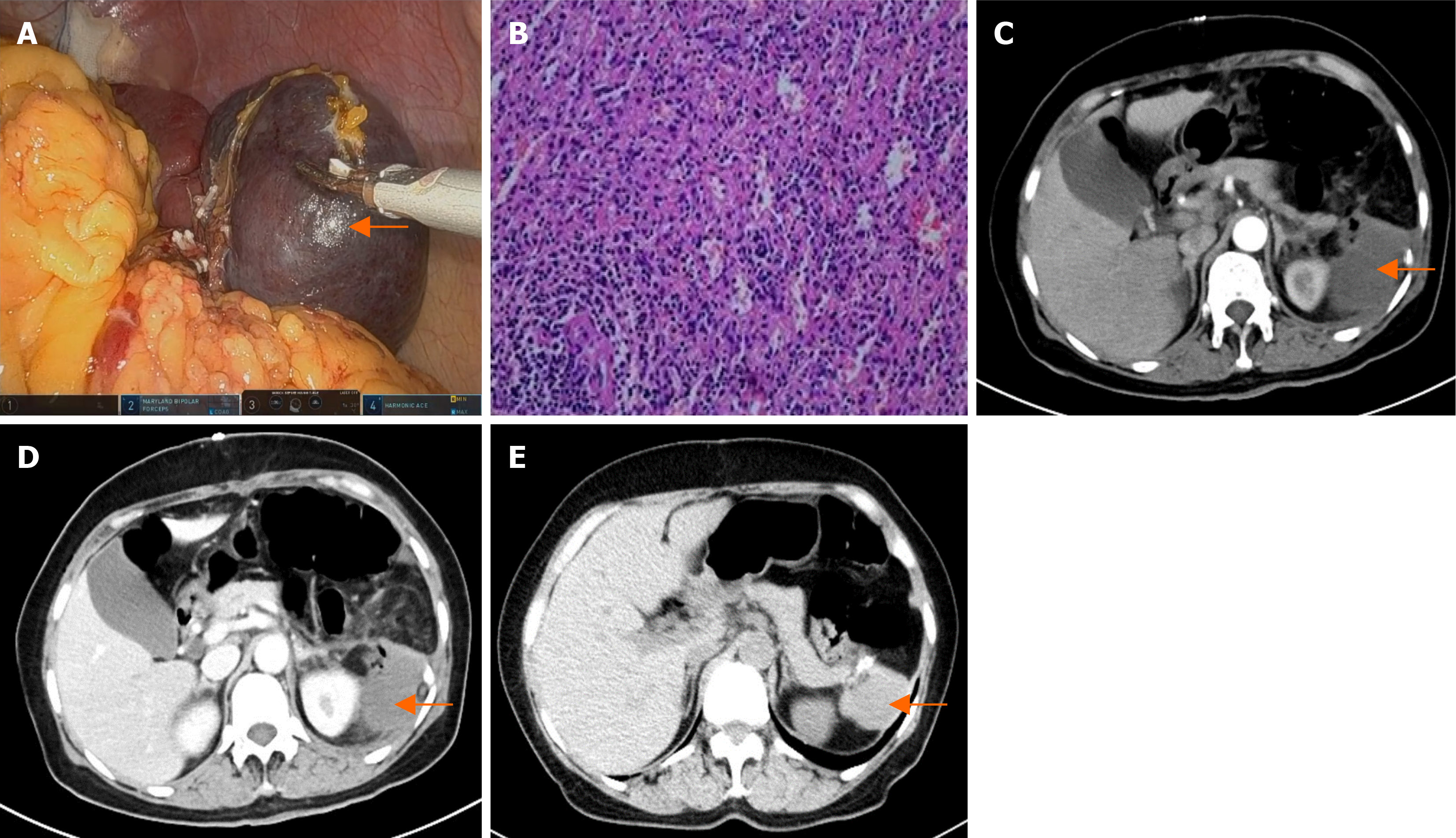
Figure 6 The patient 3's pathology result and postoperative computed tomography images.
A: Resected specimen from patient 3; B: Histopathological analysis and immunohistochemical examination of the resected specimen: Splenic hemangiomas; C and D: Computed tomography (CT) images of patient 3 on the third day after surgery. There was some effusion above the spleen (arrows); E and F: A follow-up CT scan of patient 3 at six months post-surgery indicated that there was no tumor in the spleen (arrows).
- Citation: Xue HM, Chen P, Zhu XJ, Jiao JY, Wang P. Robot-assisted partial splenectomy for benign splenic tumors: Four case reports. World J Clin Oncol 2024; 15(10): 1366-1375
- URL: https://www.wjgnet.com/2218-4333/full/v15/i10/1366.htm
- DOI: https://dx.doi.org/10.5306/wjco.v15.i10.1366


