Copyright
©The Author(s) 2024.
World J Clin Oncol. Oct 24, 2024; 15(10): 1366-1375
Published online Oct 24, 2024. doi: 10.5306/wjco.v15.i10.1366
Published online Oct 24, 2024. doi: 10.5306/wjco.v15.i10.1366
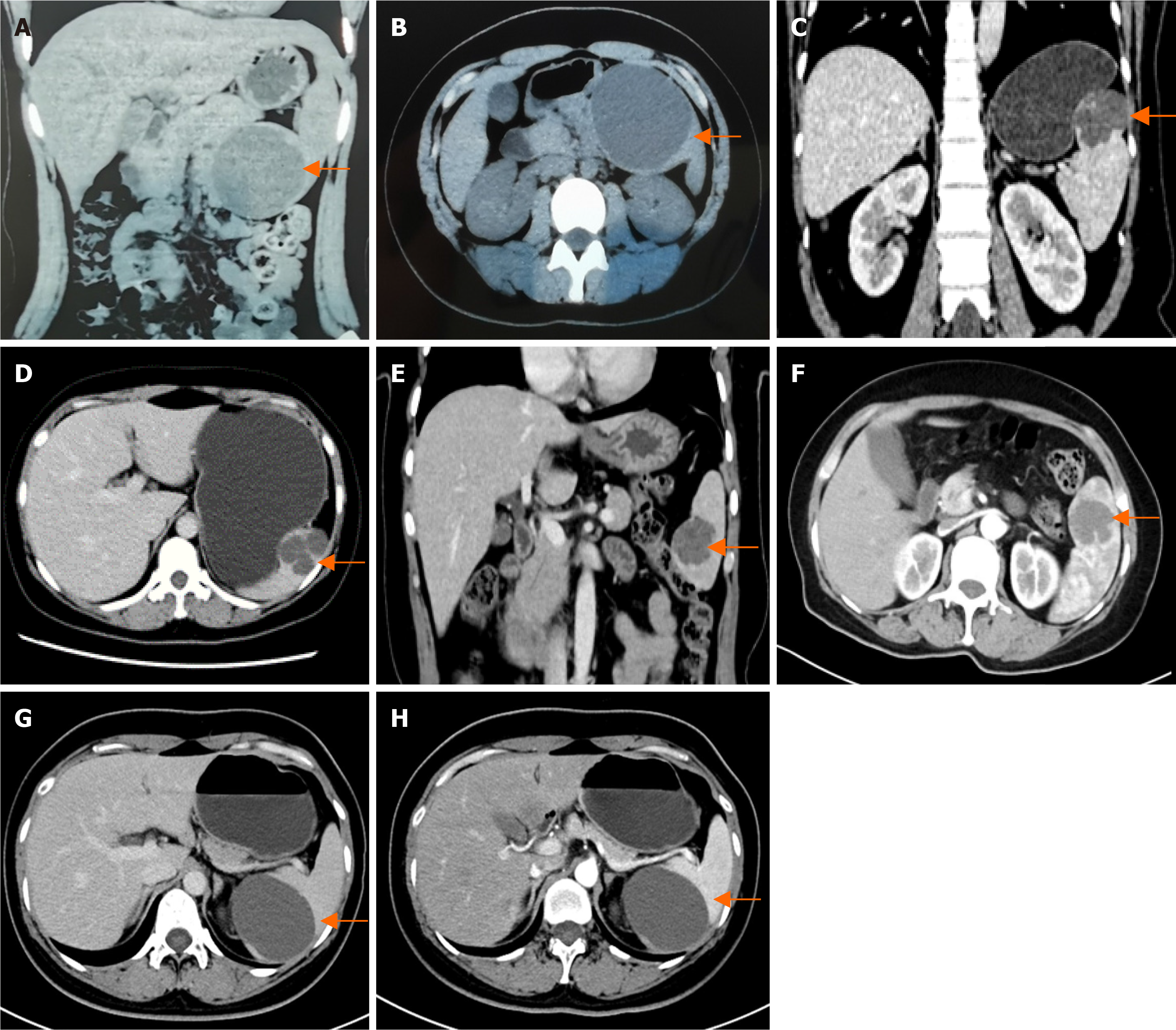
Figure 1 Imaging examinations of four patients.
A and B: Computed tomography (CT) revealed a round, non-enhancing low-density lesion measuring approximately 90 mm in the lower pole of the spleen (arrows), which was consistent with a splenic cyst; C and D: CT revealed multiple oval-shaped low-density lesions in the upper segment of the spleen (arrows), with the largest measuring approximately 52 mm; E and F: Magnetic resonance imaging revealed a 40 mm well-defined mass in the lower pole of the spleen (arrows), which appears as a low signal on T1 weight and a high signal on T2 weight; G and H: CT revealed a round, non-enhancing low-density lesion measuring approximately 73 mm in the lower pole of the spleen (arrows).
- Citation: Xue HM, Chen P, Zhu XJ, Jiao JY, Wang P. Robot-assisted partial splenectomy for benign splenic tumors: Four case reports. World J Clin Oncol 2024; 15(10): 1366-1375
- URL: https://www.wjgnet.com/2218-4333/full/v15/i10/1366.htm
- DOI: https://dx.doi.org/10.5306/wjco.v15.i10.1366


