Copyright
©The Author(s) 2024.
World J Clin Oncol. Oct 24, 2024; 15(10): 1342-1350
Published online Oct 24, 2024. doi: 10.5306/wjco.v15.i10.1342
Published online Oct 24, 2024. doi: 10.5306/wjco.v15.i10.1342
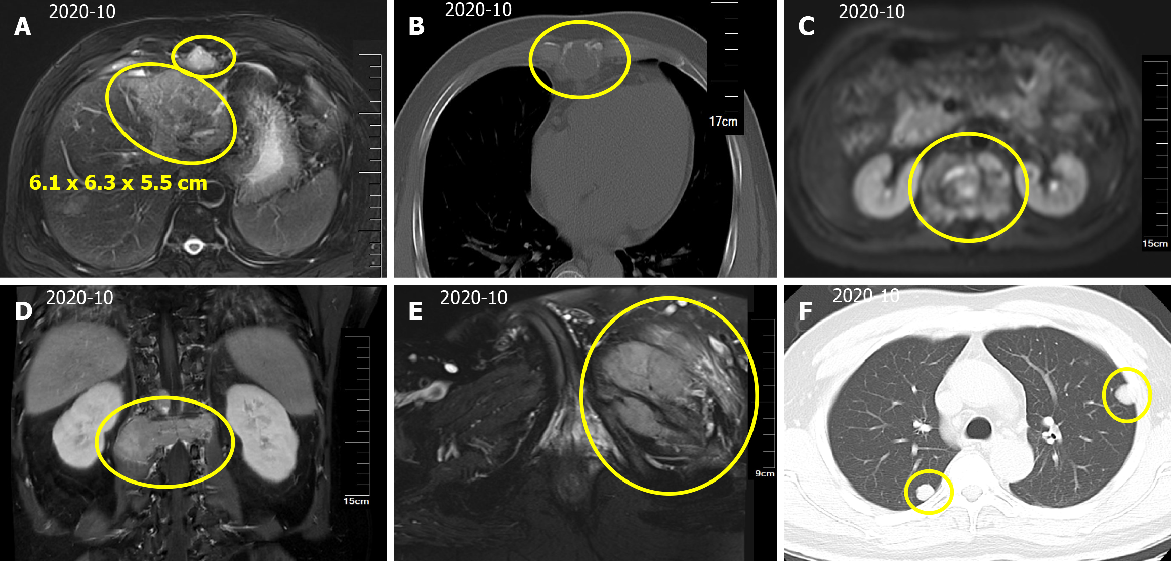
Figure 1 Hepatocellular carcinoma with intrahepatic and abdominal lymph nodes, multiple bone and lung metastases.
A: A mass in the left lobe of the liver, with a size of approximately 6.1 cm × 6.3 cm × 5.5 cm. Scattered circular abnormal signal shadows with blurred boundaries; B: Bone destruction of thoracic vertebrae and vertebral appendages; C: Paravertebral soft tissue metastases; D: L1 vertebral compression fracture, compressing spinal cord; E: Left iliac bone metastases; F: Computed tomography scan of the thorax showing multiple nodular shadows in the both lungs.
- Citation: Chen QQ, Chen CQ, Liu JK, Huang MY, Pan M, Huang H. Hypofractionated and intensity-modulated radiotherapy combined with systemic therapy in metastatic hepatocellular carcinoma: A case report. World J Clin Oncol 2024; 15(10): 1342-1350
- URL: https://www.wjgnet.com/2218-4333/full/v15/i10/1342.htm
- DOI: https://dx.doi.org/10.5306/wjco.v15.i10.1342


