Copyright
©The Author(s) 2023.
World J Clin Oncol. Apr 24, 2023; 14(4): 171-178
Published online Apr 24, 2023. doi: 10.5306/wjco.v14.i4.171
Published online Apr 24, 2023. doi: 10.5306/wjco.v14.i4.171
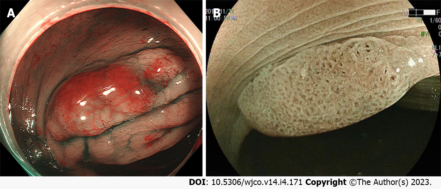
Figure 4 Endoscopic features of colorectal sessile serrated lesion cases under narrow band imaging mode.
A: The mucus cap of sessile serrated lesions (SSLs) shows a brick-red appearance under narrow band imaging (NBI); B: The expansion of the surface crypt in the SSLs shows a black spot under NBI.
- Citation: Wang RG, Wei L, Jiang B. Current progress on the endoscopic features of colorectal sessile serrated lesions. World J Clin Oncol 2023; 14(4): 171-178
- URL: https://www.wjgnet.com/2218-4333/full/v14/i4/171.htm
- DOI: https://dx.doi.org/10.5306/wjco.v14.i4.171


