Copyright
©The Author(s) 2023.
World J Clin Oncol. Apr 24, 2023; 14(4): 171-178
Published online Apr 24, 2023. doi: 10.5306/wjco.v14.i4.171
Published online Apr 24, 2023. doi: 10.5306/wjco.v14.i4.171
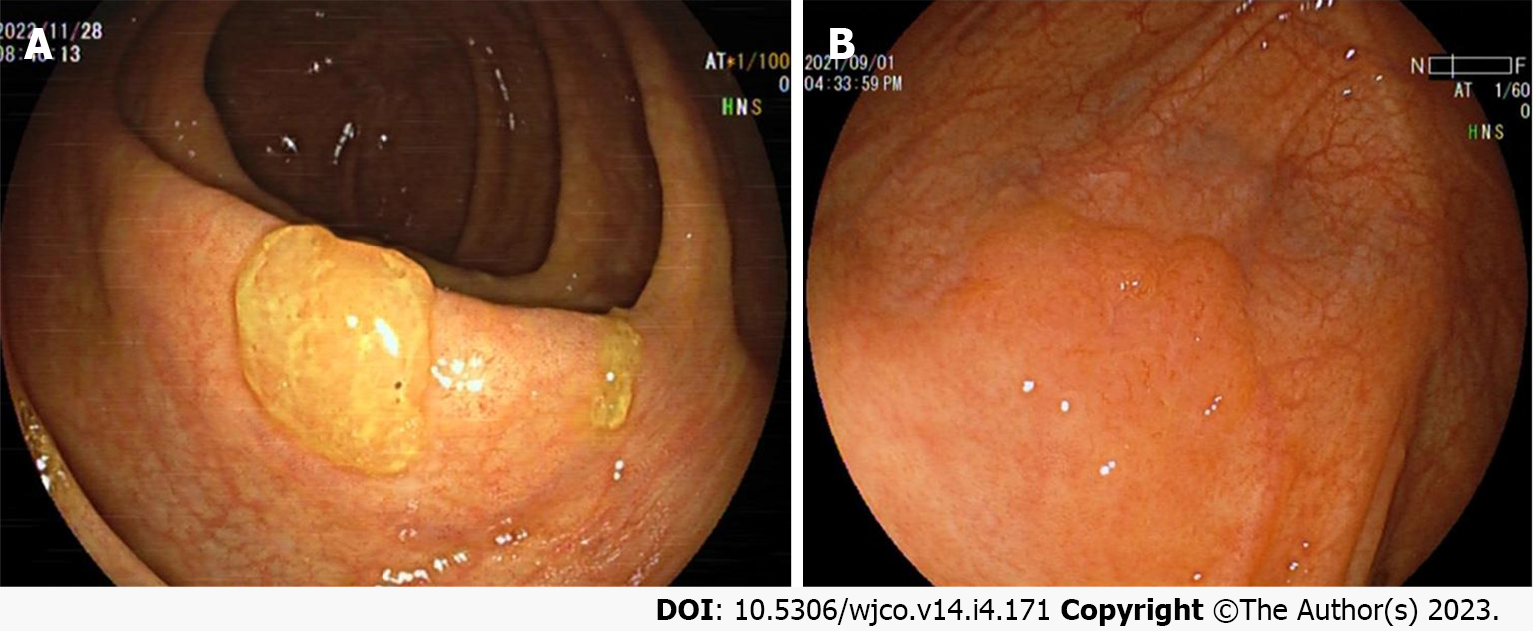
Figure 1 White light endoscopic features of colorectal sessile serrated lesion cases.
A: The sessile serrated lesions (SSLs) case with mucus cap under white light endoscopy; B: The borders of SSLs are not clearly distinguishable from the surrounding mucosa, and the morphology are cloud-like surface under white light. The above figure shows a case of SSL-D which has a reddish surface and a central depression.
- Citation: Wang RG, Wei L, Jiang B. Current progress on the endoscopic features of colorectal sessile serrated lesions. World J Clin Oncol 2023; 14(4): 171-178
- URL: https://www.wjgnet.com/2218-4333/full/v14/i4/171.htm
- DOI: https://dx.doi.org/10.5306/wjco.v14.i4.171


