Copyright
©The Author(s) 2023.
World J Clin Oncol. Dec 24, 2023; 14(12): 620-627
Published online Dec 24, 2023. doi: 10.5306/wjco.v14.i12.620
Published online Dec 24, 2023. doi: 10.5306/wjco.v14.i12.620
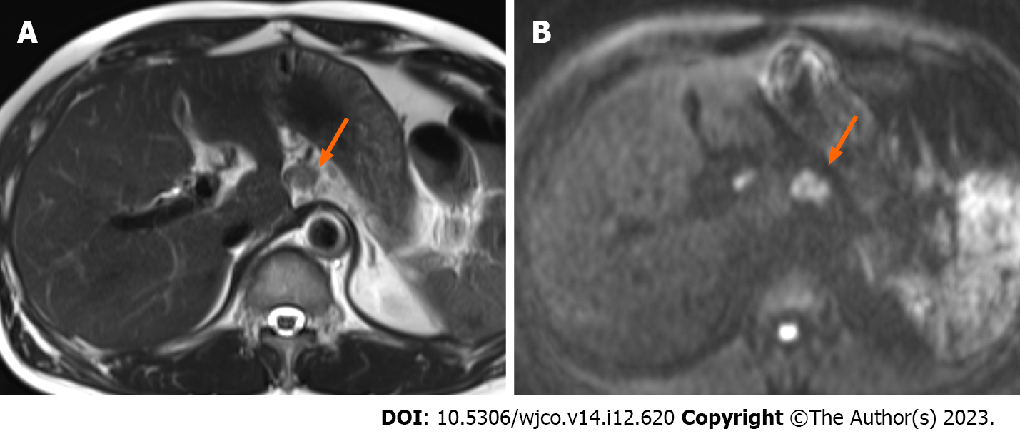
Figure 5 Magnetic resonance imaging 1 year and 10 mo postoperatively.
A: T2-weighted magnetic resonance imaging showed a small nodule in the postoperative area; B: Diffusion-weighted imaging showed a high signal in the same area, indicating recurrence in the retroperitoneal lymph nodes, arrow.
- Citation: Yamamoto K, Takada Y, Kobayashi T, Ito R, Ikeda Y, Ota S, Adachi K, Shimada Y, Hayashi M, Itani T, Asai S, Nakamura K. Rapid transformation of branched pancreatic duct-derived intraductal tubulopapillary neoplasm into an invasive carcinoma: A case report. World J Clin Oncol 2023; 14(12): 620-627
- URL: https://www.wjgnet.com/2218-4333/full/v14/i12/620.htm
- DOI: https://dx.doi.org/10.5306/wjco.v14.i12.620


