Copyright
©The Author(s) 2022.
World J Clin Oncol. May 24, 2022; 13(5): 323-338
Published online May 24, 2022. doi: 10.5306/wjco.v13.i5.323
Published online May 24, 2022. doi: 10.5306/wjco.v13.i5.323
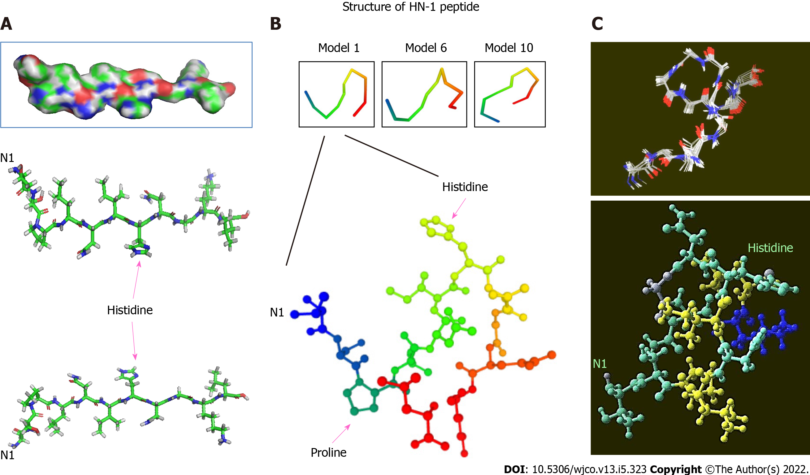
Figure 1 Three-dimensional structure of HN-1 peptide.
A: A 3D model of HN-1 peptide generated using PyMol molecular graphics system, version 1.2r3pre, Schrödinger, LLC. All graphics depict an identical configuration with the bottom two panels in the opposite orientation; B: An ensemble of de novo conformations generated by PEP-FOLD (INSERM, France) in RPBS bioinformatics web portal: https://bioserv.rpbs.univ-paris-diderot.fr/services/PEP-FOLD/; C: A 3-dimensional profile of the lowest energy structure obtained for HN-1 peptide was viewed using Raswin computer modeling software. All structures were generated using TSPLNIHNGQKL as the raw input peptide sequence. N1: The N-terminal residue.
- Citation: Hong FU, Castro M, Linse K. Tumor specifically internalizing peptide ‘HN-1’: Targeting the putative receptor retinoblastoma-regulated discoidin domain receptor 1 involved in metastasis. World J Clin Oncol 2022; 13(5): 323-338
- URL: https://www.wjgnet.com/2218-4333/full/v13/i5/323.htm
- DOI: https://dx.doi.org/10.5306/wjco.v13.i5.323


