Copyright
©The Author(s) 2022.
World J Clin Oncol. Mar 24, 2022; 13(3): 168-185
Published online Mar 24, 2022. doi: 10.5306/wjco.v13.i3.168
Published online Mar 24, 2022. doi: 10.5306/wjco.v13.i3.168
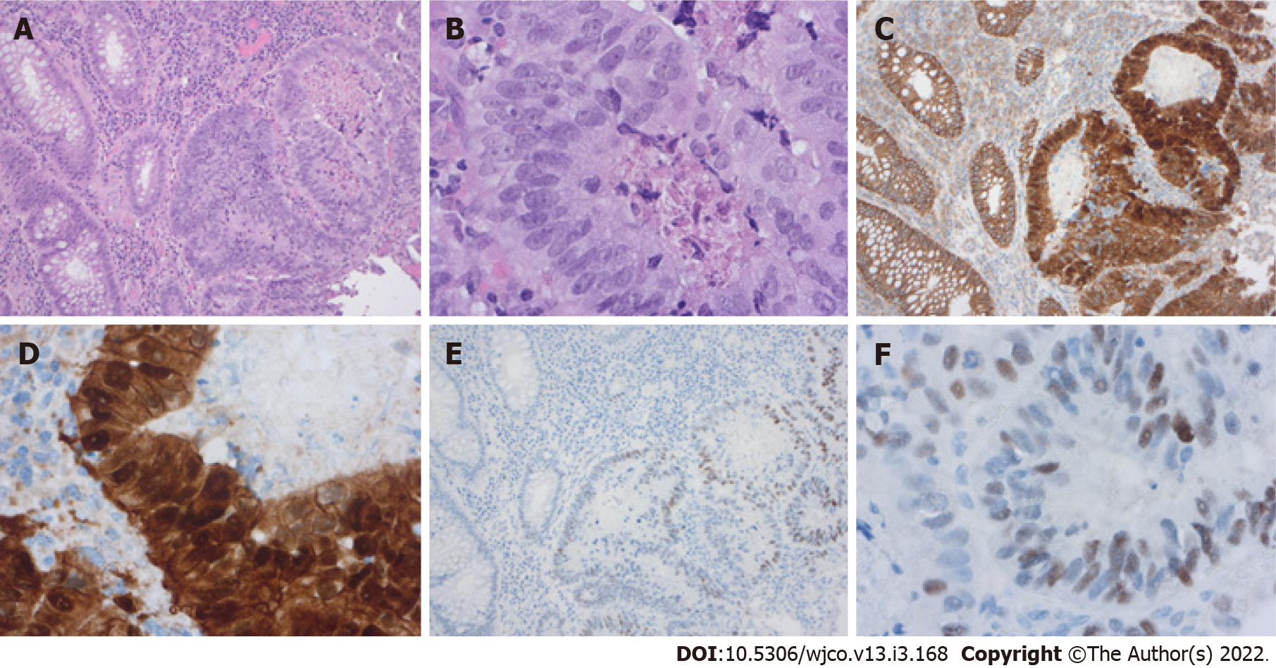
Figure 4 Colorectal carcinoma.
A: Invasive growth and loss of polarity [100 × Hematoxylin eosin (HE)]; B: Cellular atypies (400 × HE); C: β-catenin staining (100 ×) membranous in normal epithelial, nuclear staining in dysplastic cells; D: β-catenin staining (400 ×) with partly extensive accumulation of β-catenin in the nucleus; E: Positive staining of c-myc (a target of β-catenin) in the dysplastic cells (100 ×); F: Positive nuclear staining of c-myc (400 ×).
- Citation: Swoboda J, Mittelsdorf P, Chen Y, Weiskirchen R, Stallhofer J, Schüle S, Gassler N. Intestinal Wnt in the transition from physiology to oncology. World J Clin Oncol 2022; 13(3): 168-185
- URL: https://www.wjgnet.com/2218-4333/full/v13/i3/168.htm
- DOI: https://dx.doi.org/10.5306/wjco.v13.i3.168


