Copyright
©The Author(s) 2022.
World J Clin Oncol. Nov 24, 2022; 13(11): 880-895
Published online Nov 24, 2022. doi: 10.5306/wjco.v13.i11.880
Published online Nov 24, 2022. doi: 10.5306/wjco.v13.i11.880
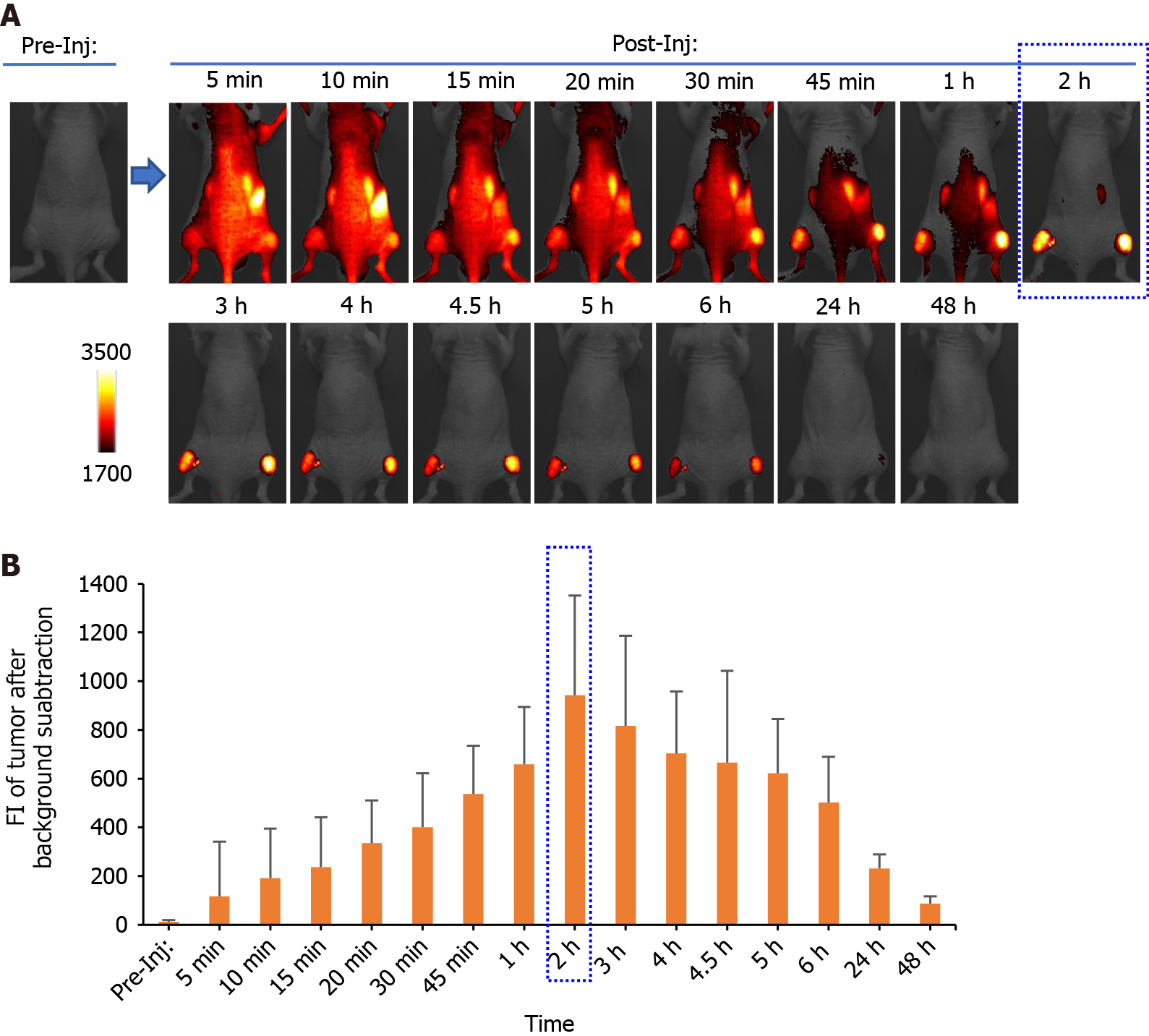
Figure 4 In vivo longitudinal near-infrared fluorescence imaging of folate-Si-rhodamine-1 in a KB tumor-bearing representative mouse.
A: Imaging was conducted before and after folate-Si-rhodamine-1 (FolateSiR-1) injection. The indicated time points after injection are at 5 min, 10 min, 15 min, 20 min, 30 min, 45 min, 1 h, 2 h, 3 h, 4 h, 4.5 h, 5 h, 6 h, 24 h, and 48 h. Time-dependent FolateSiR-1 distribution and its specific tumor accumulation is shown in serial images; B: Quantitative fluorescent signal intensity analysis of the tumors. Data are presented as the mean ± SD (n = 4). NIR: Near-infrared; FI: Fluorescent signal intensity; Pre-Inj: Pre-injection; Post-Inj: Post-injection.
- Citation: Aung W, Tsuji AB, Hanaoka K, Higashi T. Folate receptor-targeted near-infrared photodynamic therapy for folate receptor-overexpressing tumors. World J Clin Oncol 2022; 13(11): 880-895
- URL: https://www.wjgnet.com/2218-4333/full/v13/i11/880.htm
- DOI: https://dx.doi.org/10.5306/wjco.v13.i11.880


