Copyright
©The Author(s) 2021.
World J Clin Oncol. Aug 24, 2021; 12(8): 688-701
Published online Aug 24, 2021. doi: 10.5306/wjco.v12.i8.688
Published online Aug 24, 2021. doi: 10.5306/wjco.v12.i8.688
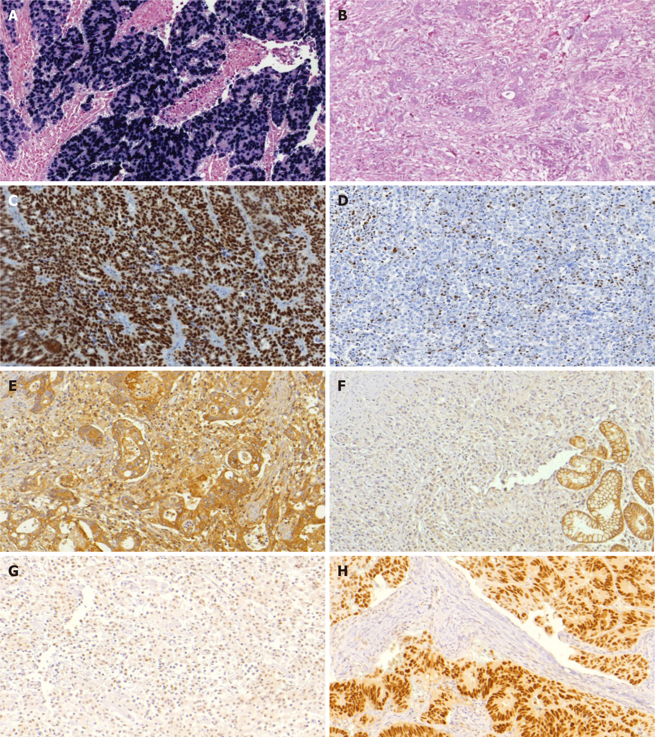
Figure 2 Microscopic findings in gastric cancer cases.
A: Tumor positive for Epstein-Barr virus (EBV) by in situ hybridization; B: Gastric cancer (GC) negative for EBV infection; C: Imuno-histoquímico (IHC) analysis of MLH1 expression in tumors with retained expression of MLH1; D: Loss of MLH1 expression; E: GC with preserved E-cadherin expression; F: tumor exhibiting loss of E-cadherin expression; G: Normal IHC expression of p53; H: tumor with aberrant p53 expression.
- Citation: Ramos MFKP, Pereira MA, de Mello ES, Cirqueira CDS, Zilberstein B, Alves VAF, Ribeiro-Junior U, Cecconello I. Gastric cancer molecular classification based on immunohistochemistry and in situ hybridization: Analysis in western patients after curative-intent surgery. World J Clin Oncol 2021; 12(8): 688-701
- URL: https://www.wjgnet.com/2218-4333/full/v12/i8/688.htm
- DOI: https://dx.doi.org/10.5306/wjco.v12.i8.688


