Copyright
©The Author(s) 2021.
World J Clin Oncol. May 24, 2021; 12(5): 323-334
Published online May 24, 2021. doi: 10.5306/wjco.v12.i5.323
Published online May 24, 2021. doi: 10.5306/wjco.v12.i5.323
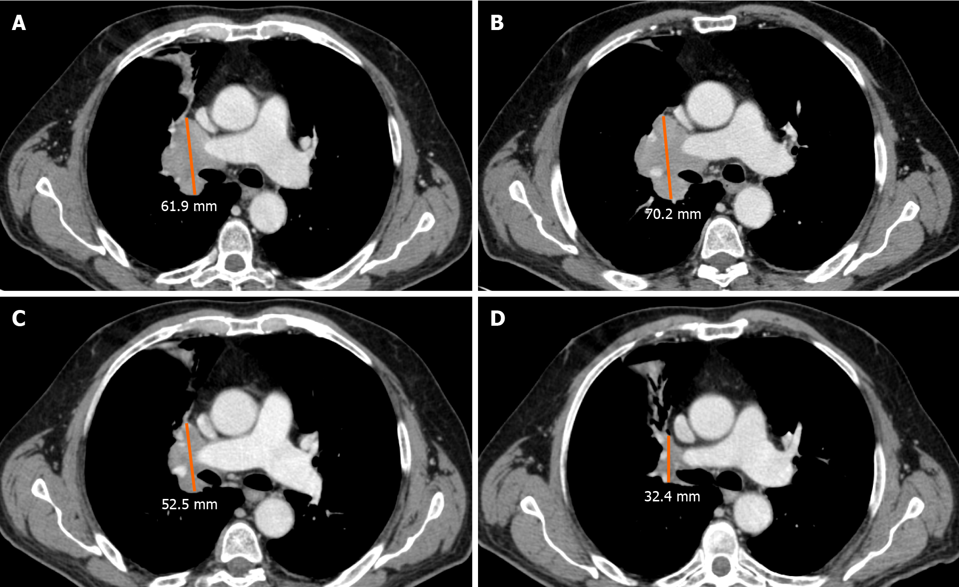
Figure 2 Axial computed tomography images in the portal-venous phase of a 69 y/o male, ex-smoker with non-small lung cell carcinoma, during second-line therapy with Atezolizumab.
A: Pre-treatment imaging show the right peri-hilar lesion; B: During follow-up after 4 wk the lesion increase in size; C and D: During the following computed tomography scans (8 and 12 wk) a significant decrease in longest diameter was achieved, confirming a final response to treatment with the presence of intercurrent (B) pseudoprogression.
- Citation: Ippolito D, Maino C, Ragusi M, Porta M, Gandola D, Franzesi CT, Giandola TP, Sironi S. Immune response evaluation criteria in solid tumors for assessment of atypical responses after immunotherapy. World J Clin Oncol 2021; 12(5): 323-334
- URL: https://www.wjgnet.com/2218-4333/full/v12/i5/323.htm
- DOI: https://dx.doi.org/10.5306/wjco.v12.i5.323


