Copyright
©The Author(s) 2021.
World J Clin Oncol. Dec 24, 2021; 12(12): 1227-1243
Published online Dec 24, 2021. doi: 10.5306/wjco.v12.i12.1227
Published online Dec 24, 2021. doi: 10.5306/wjco.v12.i12.1227
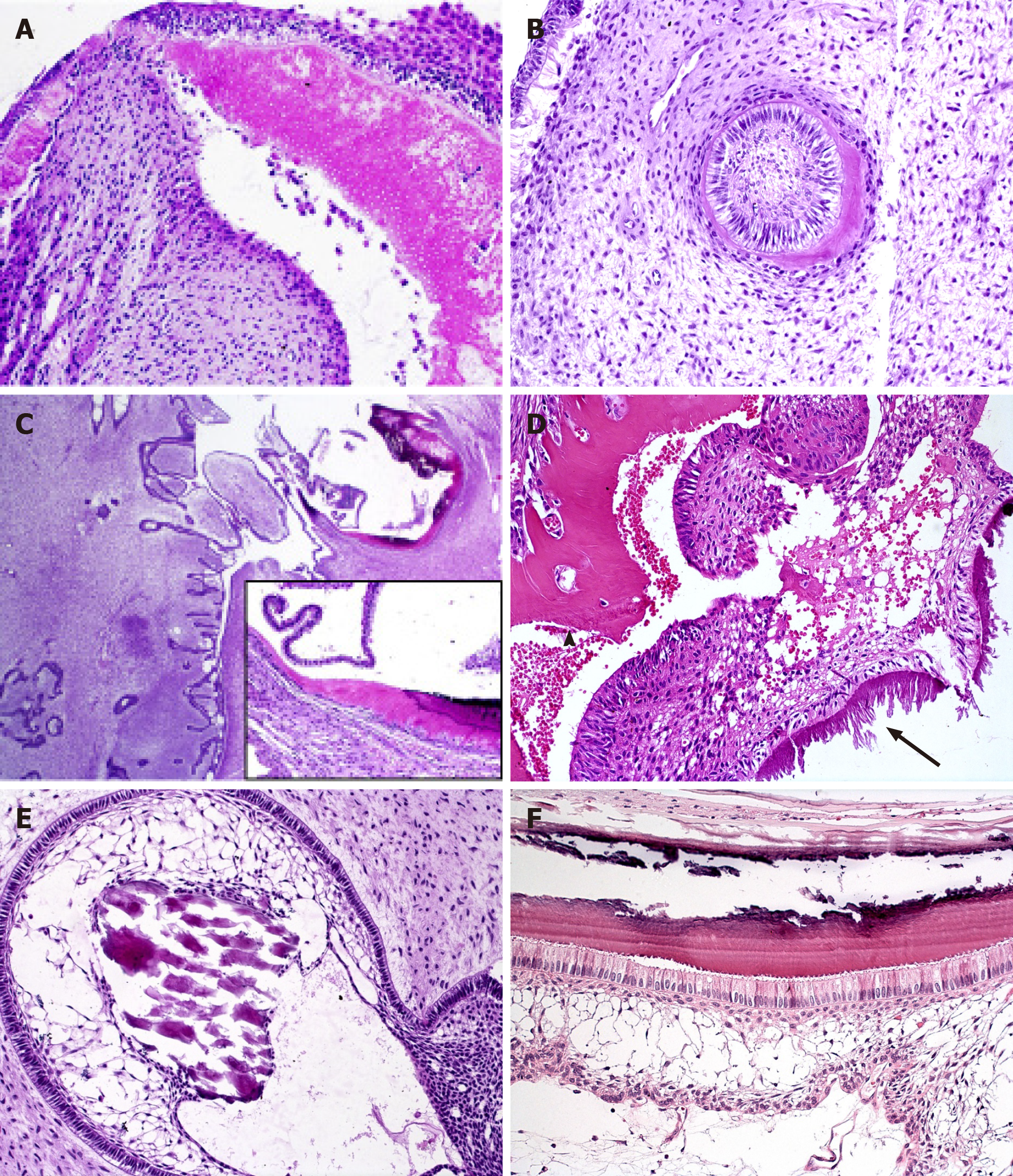
Figure 2 Mineralized tissue formation in ameloblastic fibrodentinoma and ameloblastic fibro-odontoma.
A, B: Dentinoid induction by epithelial cells in ameloblastic fibrodentinoma; note the presence of tubules in (A) (HE, 20×); C: Prominent proliferation of soft tissue similar to ameloblastic fibromas and focal areas of dentinoid and enamel matrix production in close relationship with the epithelial component in ameloblastic fibro-odontoma (HE, 2.5×; inset 20×); D: Structures similar to tubules are observed in the dentinoid (arrowhead), which can be associated with odontogenic epithelium or ectomesenchymal tissue, while enamel matrix (arrow) associated with columnar odontogenic appears more basophilic, with different patterns of deposition that can resemble prisms or globules (HE, 20×); E: Calcificated material, compatible with enameloid, in direct relationship with epithelial cells of the stellate reticulum-like area (HE, 20×); F: Details of the columnar ameloblast-like cells with reverse polarization producing enamel matrix in which the “fish scale” pattern is visible. Flattened cells between the columnar cells and stellate reticulum-like area resemble the stratum intermedium of the tooth germ, which is believed to assist the ameloblast in producing enamel during odontogenesis (HE, 40×).
- Citation: Sánchez-Romero C, Paes de Almeida O, Bologna-Molina R. Mixed odontogenic tumors: A review of the clinicopathological and molecular features and changes in the WHO classification. World J Clin Oncol 2021; 12(12): 1227-1243
- URL: https://www.wjgnet.com/2218-4333/full/v12/i12/1227.htm
- DOI: https://dx.doi.org/10.5306/wjco.v12.i12.1227


