Copyright
©The Author(s) 2021.
World J Clin Oncol. Nov 24, 2021; 12(11): 1072-1082
Published online Nov 24, 2021. doi: 10.5306/wjco.v12.i11.1072
Published online Nov 24, 2021. doi: 10.5306/wjco.v12.i11.1072
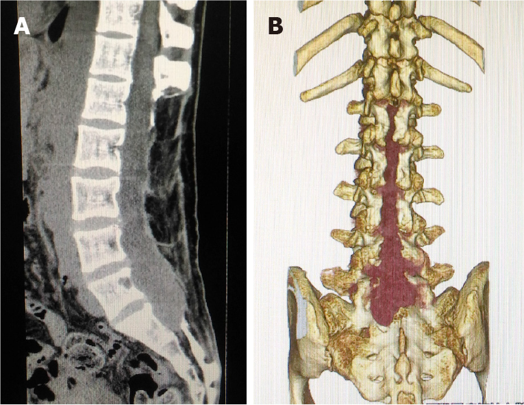
Figure 2 Computed tomography at ultra-late recurrence of a myxopapillary ependymoma.
A: A plain sagittal image demonstrated the previous laminectomy area (L1-S1); B: Three-dimensional image demonstrated a recurrent tumor (dark red) in the previous laminectomy area (L1-S1).
- Citation: Kanno H, Kanetsuna Y, Shinonaga M. Anaplastic myxopapillary ependymoma: A case report and review of literature. World J Clin Oncol 2021; 12(11): 1072-1082
- URL: https://www.wjgnet.com/2218-4333/full/v12/i11/1072.htm
- DOI: https://dx.doi.org/10.5306/wjco.v12.i11.1072


