Copyright
©The Author(s) 2021.
World J Clin Oncol. Oct 24, 2021; 12(10): 926-934
Published online Oct 24, 2021. doi: 10.5306/wjco.v12.i10.926
Published online Oct 24, 2021. doi: 10.5306/wjco.v12.i10.926
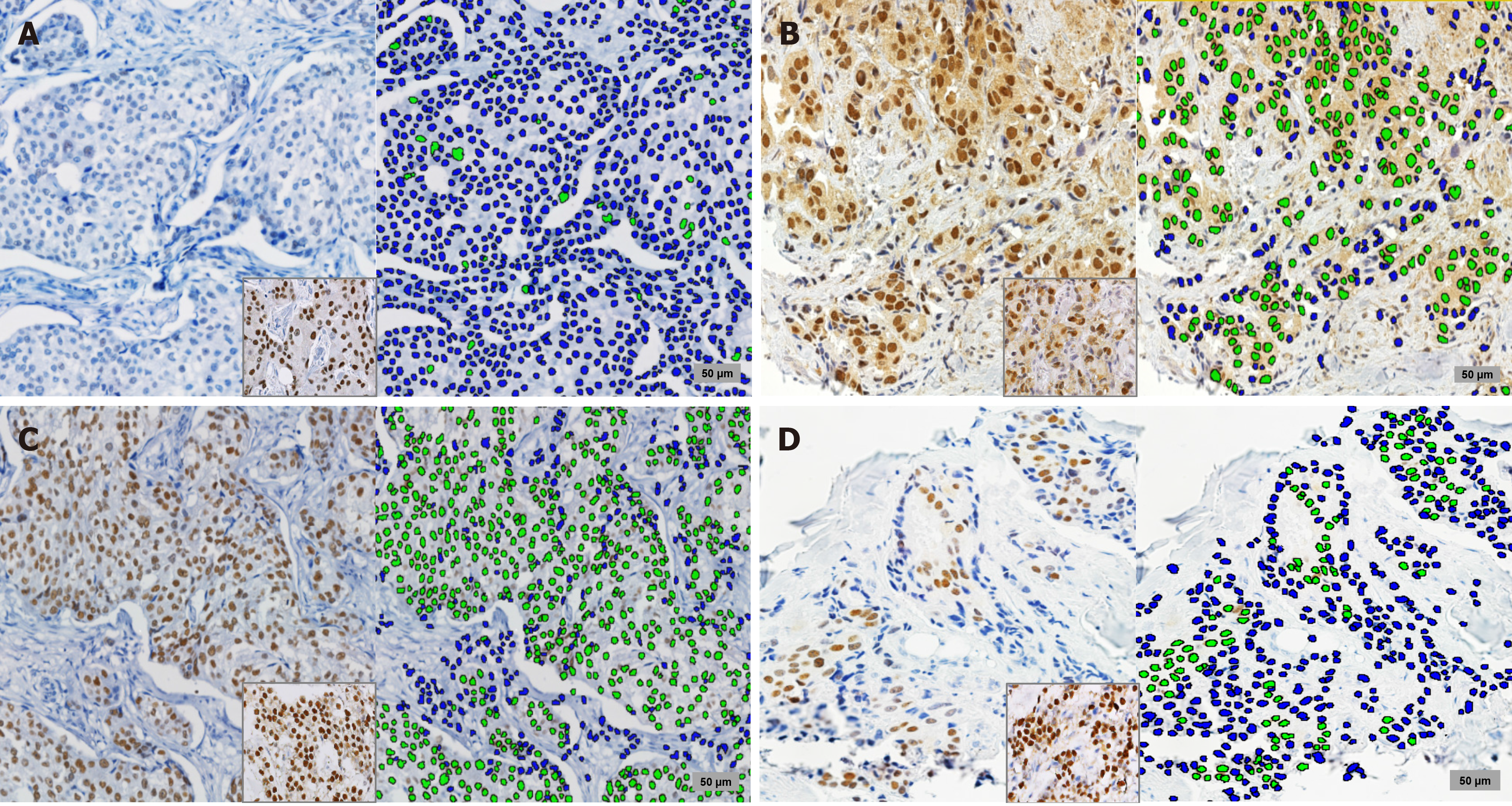
Figure 1 Immunohistochemical staining and automated scoring.
Sections show positive staining for androgen receptor (A and B) and estrogen receptor (C and D) from the same tissue areas and the positive control. In the left, sections show images of the immunohistochemical studies (× 20). In the right, the automated analysis is presented, where negative nuclei are highlighted in blue and positive nuclei in green.
- Citation: Castaneda CA, Castillo M, Bernabe LA, Sanchez J, Torres E, Suarez N, Tello K, Fuentes H, Dunstan J, De La Cruz M, Cotrina JM, Abugattas J, Guerra H, Gomez HL. A biomarker study in Peruvian males with breast cancer. World J Clin Oncol 2021; 12(10): 926-934
- URL: https://www.wjgnet.com/2218-4333/full/v12/i10/926.htm
- DOI: https://dx.doi.org/10.5306/wjco.v12.i10.926


