Copyright
©The Author(s) 2021.
World J Clin Oncol. Oct 24, 2021; 12(10): 897-911
Published online Oct 24, 2021. doi: 10.5306/wjco.v12.i10.897
Published online Oct 24, 2021. doi: 10.5306/wjco.v12.i10.897
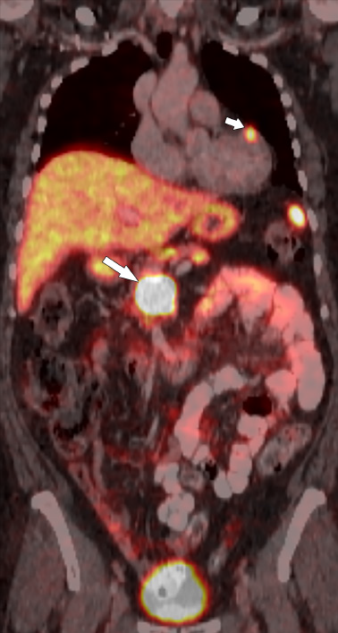
Figure 4 Sixty-two-year-old female with metastatic pancreatic neuroendocrine neoplasm.
Coronal fused Gallium-68 1,4,7,10-tetraazacyclodo
- Citation: Segaran N, Devine C, Wang M, Ganeshan D. Current update on imaging for pancreatic neuroendocrine neoplasms. World J Clin Oncol 2021; 12(10): 897-911
- URL: https://www.wjgnet.com/2218-4333/full/v12/i10/897.htm
- DOI: https://dx.doi.org/10.5306/wjco.v12.i10.897


