Copyright
©The Author(s) 2021.
World J Clin Oncol. Oct 24, 2021; 12(10): 897-911
Published online Oct 24, 2021. doi: 10.5306/wjco.v12.i10.897
Published online Oct 24, 2021. doi: 10.5306/wjco.v12.i10.897
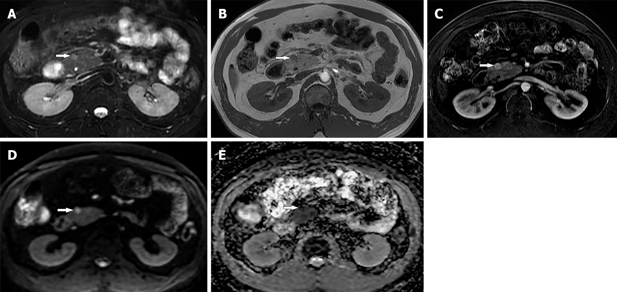
Figure 3 Thirty-five-year-old male with small pancreatic neuroendocrine neoplasm.
A: Axial magnetic resonance T2 weighted image; B: T1 weighted image show a small 1 cm mass (arrow) in the head of pancreas; C: Arterial phase image shows avid enhancement in the tumor; D: Diffusion-weighted image; E: Apparent diffusion coefficient map show restricted diffusion within the tumor (arrow). Biopsy confirmed diagnosis of pancreatic neuroendocrine neoplasm.
- Citation: Segaran N, Devine C, Wang M, Ganeshan D. Current update on imaging for pancreatic neuroendocrine neoplasms. World J Clin Oncol 2021; 12(10): 897-911
- URL: https://www.wjgnet.com/2218-4333/full/v12/i10/897.htm
- DOI: https://dx.doi.org/10.5306/wjco.v12.i10.897


