Copyright
©The Author(s) 2021.
World J Clin Oncol. Oct 24, 2021; 12(10): 897-911
Published online Oct 24, 2021. doi: 10.5306/wjco.v12.i10.897
Published online Oct 24, 2021. doi: 10.5306/wjco.v12.i10.897
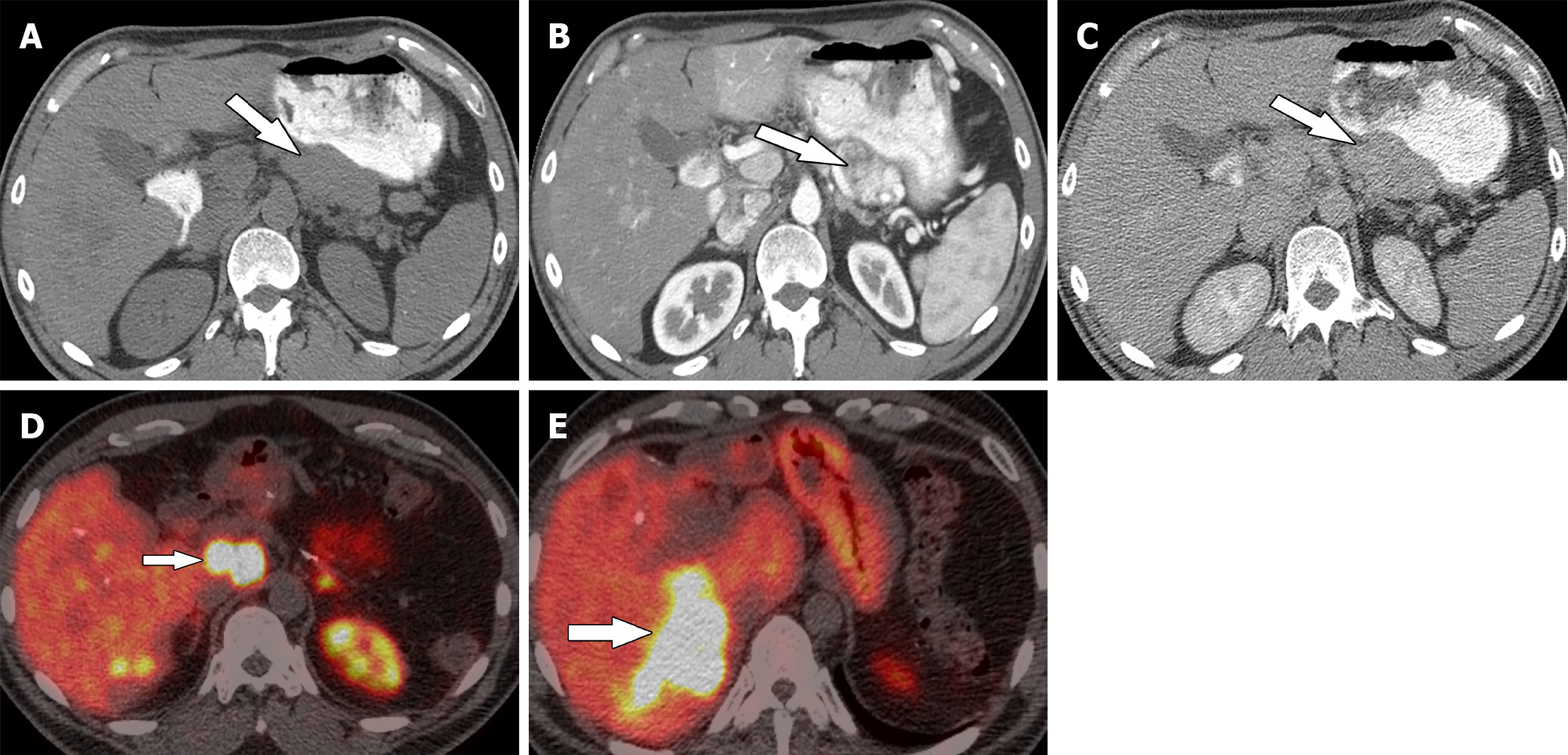
Figure 2 Thirty-eight-year-old woman with pancreatic neuroendocrine neoplasm.
A: Axial precontrast computed tomography; B and C: Contrast-enhanced computed tomography in the arterial phase (B) and delayed phase (C) demonstrate pancreatic neuroendocrine neoplasm (arrow). Patient underwent surgical resection; D and E: Follow-up Gallium-68 1,4,7,10-tetraazacyclododecane-1,4,7,10-tetraacetic acid–octreotate positron emission tomography/computed tomography shows metastatic adenopathy (short arrow) and liver metastases (long arrow).
- Citation: Segaran N, Devine C, Wang M, Ganeshan D. Current update on imaging for pancreatic neuroendocrine neoplasms. World J Clin Oncol 2021; 12(10): 897-911
- URL: https://www.wjgnet.com/2218-4333/full/v12/i10/897.htm
- DOI: https://dx.doi.org/10.5306/wjco.v12.i10.897


