Copyright
©The Author(s) 2021.
World J Clin Oncol. Oct 24, 2021; 12(10): 897-911
Published online Oct 24, 2021. doi: 10.5306/wjco.v12.i10.897
Published online Oct 24, 2021. doi: 10.5306/wjco.v12.i10.897
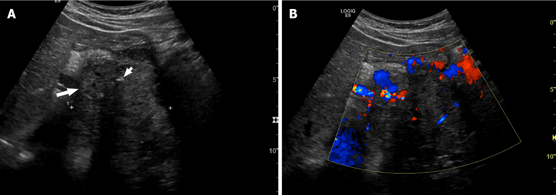
Figure 1 Forty-year-old man with pancreatic neuroendocrine neoplasm.
A: Axial ultrasound shows a large solid heterogeneous mass (long arrow). Internal calcification (small arrow) is seen, causing posterior acoustic shadowing; B: Doppler ultrasound shows increased vascularity within the pancreatic tumor.
- Citation: Segaran N, Devine C, Wang M, Ganeshan D. Current update on imaging for pancreatic neuroendocrine neoplasms. World J Clin Oncol 2021; 12(10): 897-911
- URL: https://www.wjgnet.com/2218-4333/full/v12/i10/897.htm
- DOI: https://dx.doi.org/10.5306/wjco.v12.i10.897


