Copyright
©The Author(s) 2021.
World J Clin Oncol. Jan 24, 2021; 12(1): 13-30
Published online Jan 24, 2021. doi: 10.5306/wjco.v12.i1.13
Published online Jan 24, 2021. doi: 10.5306/wjco.v12.i1.13
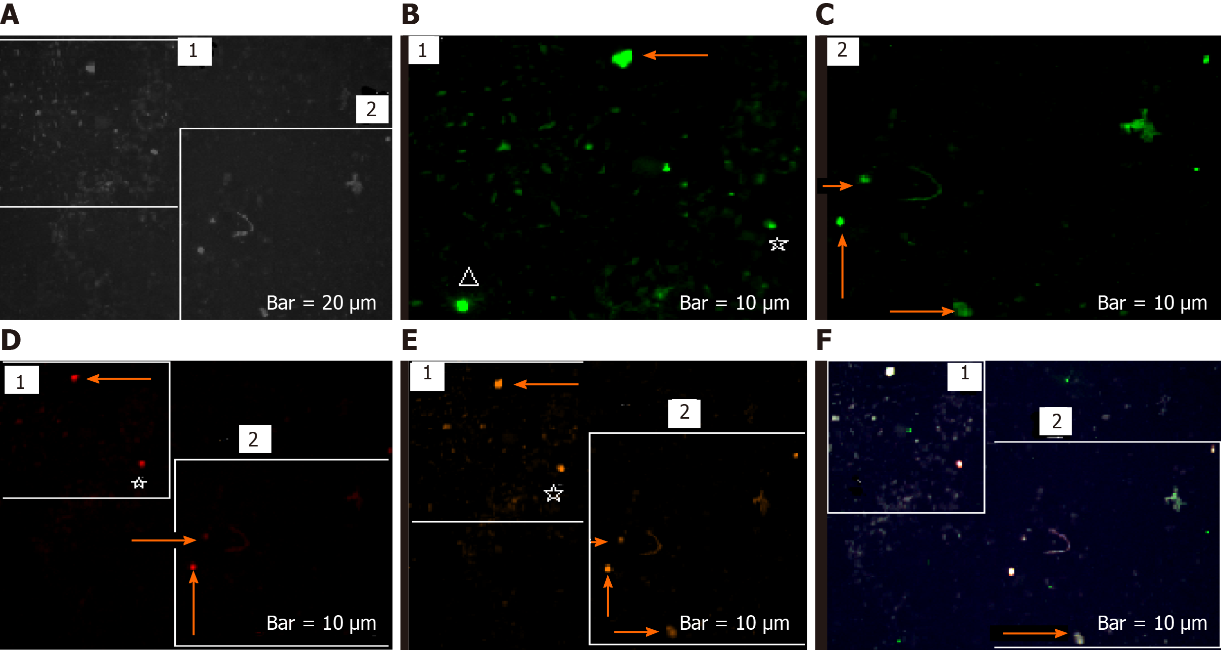
Figure 12 Status of protein expression of neuronal marker, CD133 and vascular endothelial growth factor in the brain circulating tumor cells of patient 1 affected with meningioma.
A: Circulating tumor cells (CTCs) with phase contrast; B and C: Present CTCs with Ne conjugated with fluorescein isothiocyanate; D: Present status of vascular endothelial growth factor (VEGF), conjugated with phycoerythrin–indodicarbocyanine; E: Reflect status of CD133, conjugated with R-phycoerythrin; F: Present the merged image of 4′,6-diamidino-2-phenylindole/Ne/VEGF/CD133, reflective of a remarkable co-expression between protein neuronal markers CD133 and VEGF. Arrows on the top of images B, D1, and E1 reflect more involved cells with high-, very less involved cells with much lower-, and less involved cells with high-expression for Ne, VEGF, and CD133, respectively. The asterisks on the down/right of B, B and E illustrate almost same intensity of expression, but with more CTCs involved in image B for Ne, VEGF, and CD133, respectively. A triangle symbol is reflective of a remarkable high expression for neuronal marker in CTCs. Arrows on the below/left-down of images C2, D2, and E2 present higher expression for Ne than VEGF and CD133. Arrows on the bellow/left-up of images C2, D2, and E2 illustrate low expression for Ne and CD133, and lack of expression for VEGF. The arrows down the images of C2, D2, and E2 illustrate the heterogenic (low and lack) of expression for Ne and CD133 and lack of expression for VEGF.
- Citation: Mehdipour P, Javan F, Jouibari MF, Khaleghi M, Mehrazin M. Evolutionary model of brain tumor circulating cells: Cellular galaxy. World J Clin Oncol 2021; 12(1): 13-30
- URL: https://www.wjgnet.com/2218-4333/full/v12/i1/13.htm
- DOI: https://dx.doi.org/10.5306/wjco.v12.i1.13


