Copyright
©The Author(s) 2021.
World J Clin Oncol. Jan 24, 2021; 12(1): 13-30
Published online Jan 24, 2021. doi: 10.5306/wjco.v12.i1.13
Published online Jan 24, 2021. doi: 10.5306/wjco.v12.i1.13
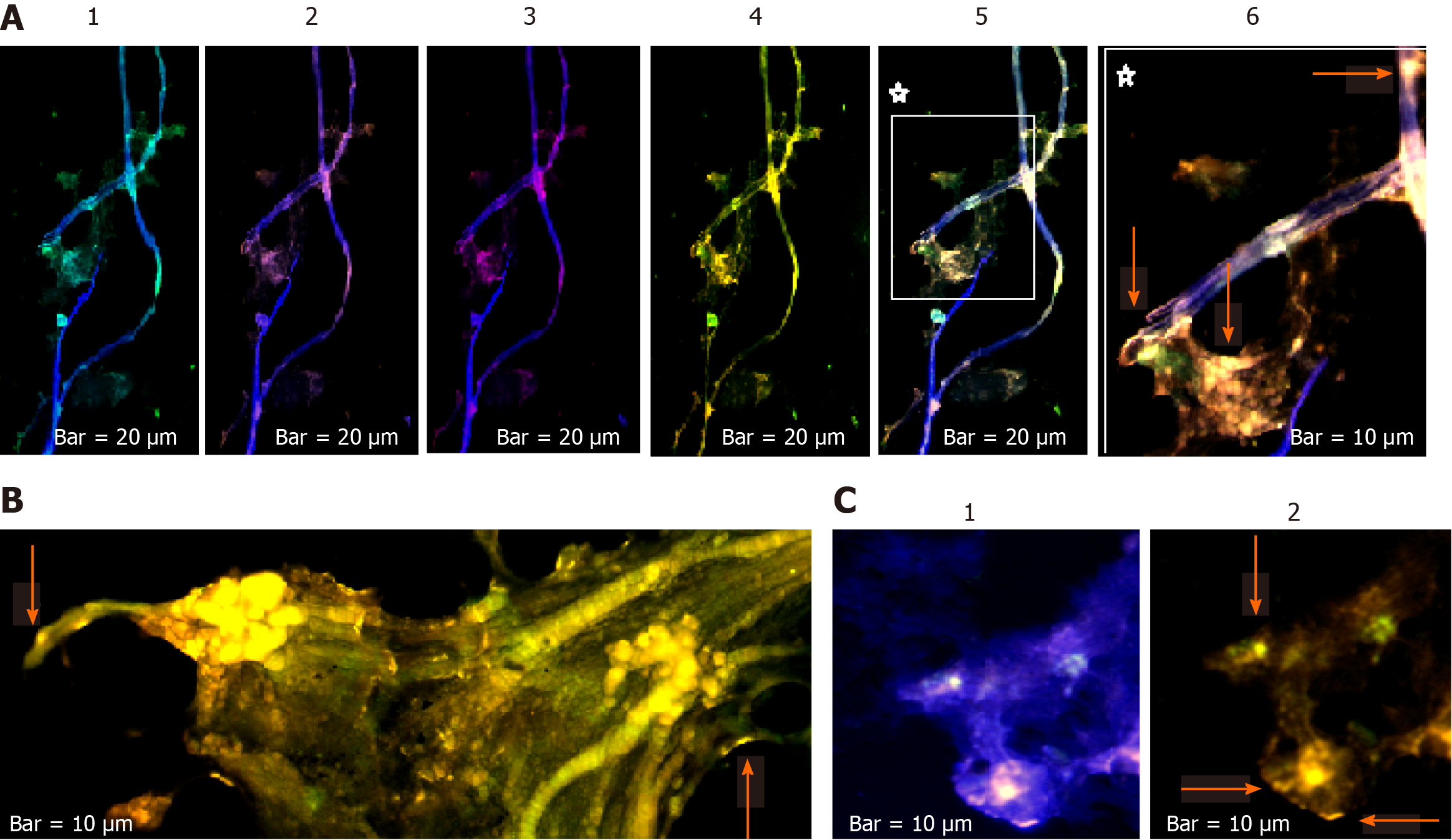
Figure 7 Protein expression status of C-C chemokine ligand 2/vascular endothelial growth factor/epidermal growth factor of two cell population from tumor sample and circulating tumor cells in patient 11 affected with meningioma.
C-C chemokine ligand 2 (CCL2), vascular endothelial growth factor (VEGF), and epidermal growth factor (EGF) are conjugated with fluorescein isothiocyanate, R-phycoerythrin, and phycoerythrin–indodicarbocyanine, respectively, in patient number 3. A: Vascular images accompanied by the partial tumor section [1: Merged of 4′,6-diamidino-2-phenylindole (DAPI)/CCL2; 2: Merged of DAPI/VEGF; 3: Merged of DAPI/ EGF; 4: Co-expression of CCL2/EGF/VEGF; 5: Co-expression of DAPI/CCL2/VEGF/EGF; 6: A cropped section of 5]; B: Image of a tumor section reflecting of the Co-expression of CCL2/VEGF/EGF; C [1:DAPI/CCL2/VEGF/EGF; 2:CCL2/VEGF/EGF]: Illustrates the CTCs in the blood sample of the same patient. The arrows are indicative of expression status in tumor cells, in vasculature, and the microvesicles.
- Citation: Mehdipour P, Javan F, Jouibari MF, Khaleghi M, Mehrazin M. Evolutionary model of brain tumor circulating cells: Cellular galaxy. World J Clin Oncol 2021; 12(1): 13-30
- URL: https://www.wjgnet.com/2218-4333/full/v12/i1/13.htm
- DOI: https://dx.doi.org/10.5306/wjco.v12.i1.13


