Copyright
©The Author(s) 2021.
World J Clin Oncol. Jan 24, 2021; 12(1): 13-30
Published online Jan 24, 2021. doi: 10.5306/wjco.v12.i1.13
Published online Jan 24, 2021. doi: 10.5306/wjco.v12.i1.13
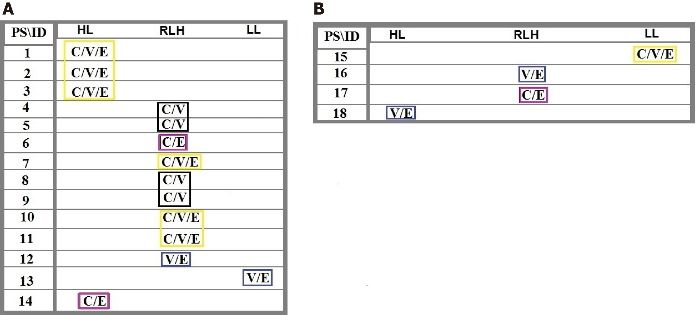
Figure 2 Ratio distribution of the cells lacking any protein expression of C-C chemokine ligand 2, vascular endothelial growth factor and epidermal growth factor between tumor cells in tumor sample to the circulated brain tumor cells in the blood.
Patients 1-14: Are affected with meningioma and patients 15-18 include 3 metastatic carcinomas (15: Brain/lung-origin; 16 and 17: Brain/breast-origin) and 1 with cerebellar medulloblastoma (CM). A: Includes patients 1-14 affected with primary meningioma; B: Includes patients 15-18 affected with the metastatic carcinoma and CM. Harmonic ratio of tumor cells to the circulated brain tumor cells are marked as specific colored quadrants related to the intensity shared classification based between different proteins. PS: Protein status; HL: Highest level; RHL: Relatively high level; LL: Low level; ID: Patients’ identification.
- Citation: Mehdipour P, Javan F, Jouibari MF, Khaleghi M, Mehrazin M. Evolutionary model of brain tumor circulating cells: Cellular galaxy. World J Clin Oncol 2021; 12(1): 13-30
- URL: https://www.wjgnet.com/2218-4333/full/v12/i1/13.htm
- DOI: https://dx.doi.org/10.5306/wjco.v12.i1.13


