Copyright
©The Author(s) 2021.
World J Clin Oncol. Jan 24, 2021; 12(1): 13-30
Published online Jan 24, 2021. doi: 10.5306/wjco.v12.i1.13
Published online Jan 24, 2021. doi: 10.5306/wjco.v12.i1.13
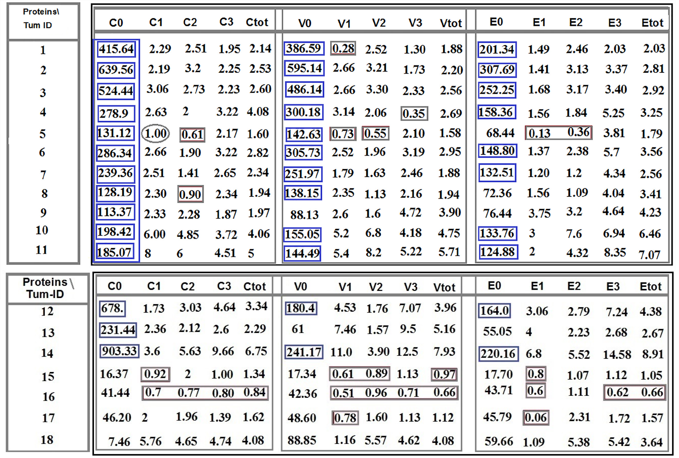
Figure 1 Ratio distribution of the cells lacking- and the classified positive-protein expression of C-C chemokine ligand 2, vascular endothelial growth factor, and epidermal growth factor between tumor cells in tumor sample to the circulated brain tumor cells.
Patients 1-11: Are affected with meningioma. Patients 12-14: Are affected with meningioma, and patients 15-18 include three metastatic carcinomas (15: Brain/lung-origin; 16 and 17: Brain breast-origin) and one with cerebellar medulloblastoma. Provided horizontal and vertical indices are related to: C0, V0, and E0: Ratio related to lack of protein expression in C-C chemokine ligand 2 (CCL2), vascular endothelial growth factor (VEGF), and epidermal growth factor (EGF), respectively; C1, V1 and E1: Low expression of CCL2, VEGF, and EGF respectively; C2, V2, and E2: Medium protein expression of CCL2, VEGF, and EGF, respectively; C3, V3, and E3: High expression of CCL2, VEGF, and EGF, respectively; Ctot, Vtot2, and Etot2: Total ratio is related to the low plus medium, plus high protein expression of CCL2, VEGF, and EGF, respectively. C: C-C chemokine ligand 2; V: Vascular endothelial growth factor; E: Epidermal growth factor.
- Citation: Mehdipour P, Javan F, Jouibari MF, Khaleghi M, Mehrazin M. Evolutionary model of brain tumor circulating cells: Cellular galaxy. World J Clin Oncol 2021; 12(1): 13-30
- URL: https://www.wjgnet.com/2218-4333/full/v12/i1/13.htm
- DOI: https://dx.doi.org/10.5306/wjco.v12.i1.13


