Copyright
©The Author(s) 2020.
World J Clin Oncol. Aug 24, 2020; 11(8): 655-672
Published online Aug 24, 2020. doi: 10.5306/wjco.v11.i8.655
Published online Aug 24, 2020. doi: 10.5306/wjco.v11.i8.655
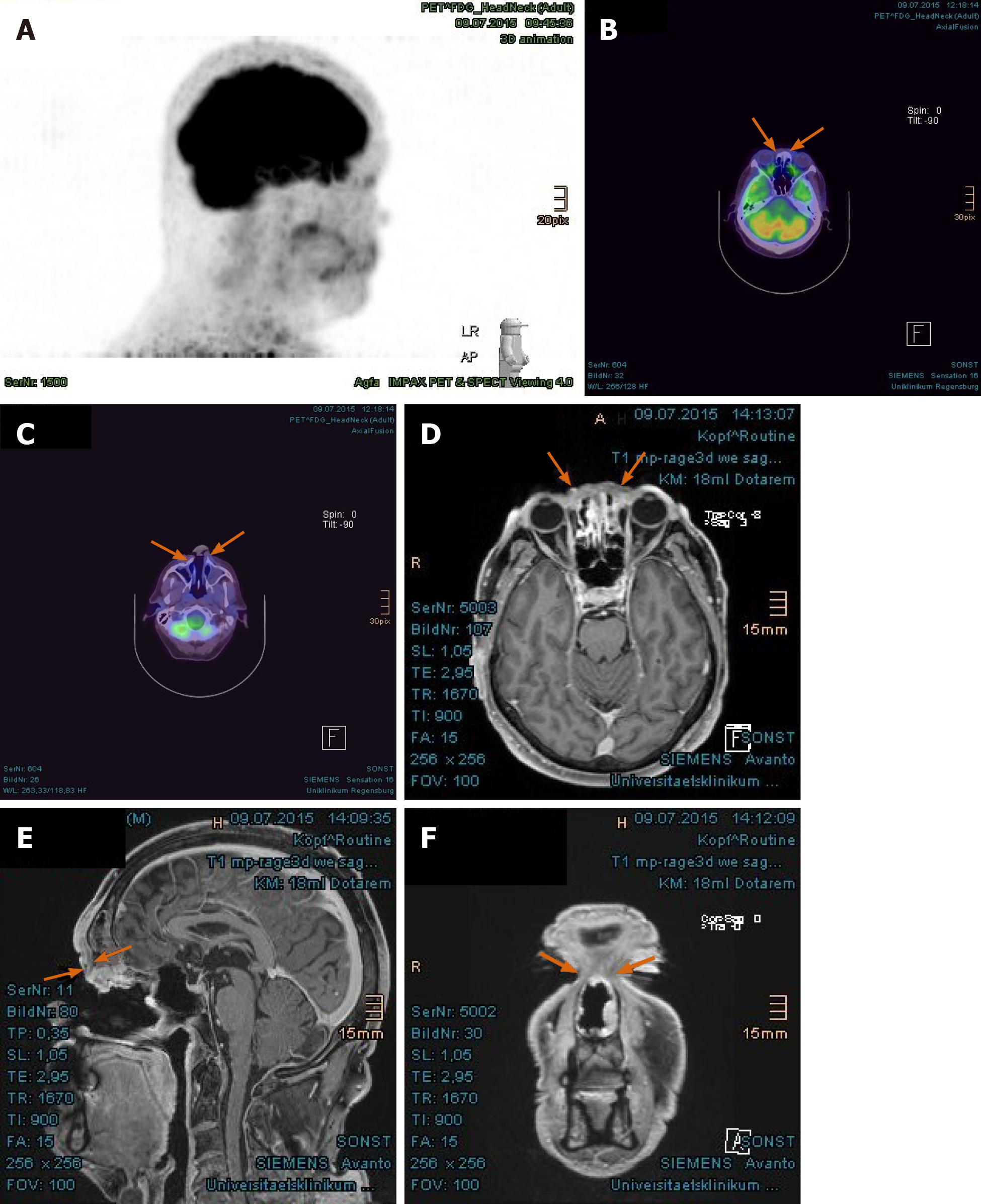
Figure 6 Complete tumor remission demonstrated in the positron emission tomography with 2-deoxy-2--fluorine-18-fluoro-D-glucose/computed tomography and magnetic resonance imaging at 8 mo after proton beam therapy.
A: No pathologically increased activity in the positron emission tomography, overview image; B: Absence of increased fluoro-D-glucose avidity in the nasal bridge (arrow-marked in positron emission tomography/computed tomography); C: Absence of metabolically active tumor in the nostrils; D: Corresponding area in the nasal bridge in magnetic resonance imaging, presented in axial plane; E: Corresponding area in the frontal skull base, presented in sagittal plane; F: Coronal presentation of tumor remission in the nasal bridge. Previously enhancing tumors marked with arrows.
- Citation: Lin YL. Proton beam therapy of periorbital sinonasal squamous cell carcinoma: Two case reports and review of literature. World J Clin Oncol 2020; 11(8): 655-672
- URL: https://www.wjgnet.com/2218-4333/full/v11/i8/655.htm
- DOI: https://dx.doi.org/10.5306/wjco.v11.i8.655


