Copyright
©The Author(s) 2020.
World J Clin Oncol. Aug 24, 2020; 11(8): 614-628
Published online Aug 24, 2020. doi: 10.5306/wjco.v11.i8.614
Published online Aug 24, 2020. doi: 10.5306/wjco.v11.i8.614
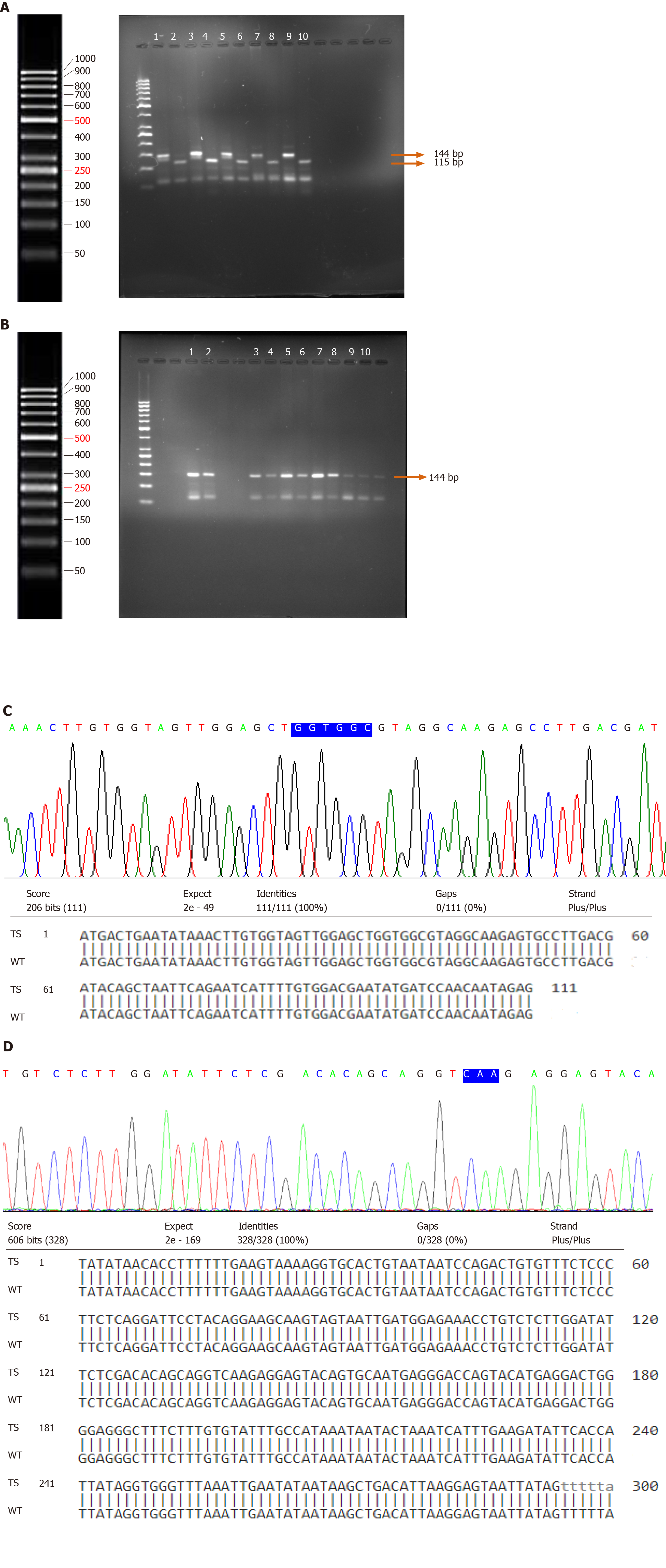
Figure 3 K-Ras gene point mutation analysis in patients diagnosed with urothelial bladder cancer.
A: Polymerase chain reaction (PCR) - restriction fragment length polymorphism (RFLP) of codon 12. Undigested amplified PCR product (144-bp); BstI-cut PCR product (115-bp and 29-bp); Lanes 1, 3, 5, 7 and 9: Undigested products and Lanes 2, 4, 6, 8 and 10: Digested PCR products. Lanes 1 and 2 represent the band patterns in normal bladder tissues whereas lanes 3, 4; 5, 6; 7, 8; and 9, 10 represent the band patterns in tumor specimens; B: PCR-RFLP of K-Ras codon 13. Undigested amplified PCR product (144-bp); Hph I-cut PCR product (101-bp and 43-bp); Lanes 1, 3, 5, 7, 9, and 11: Undigested products and Lanes 2, 4, 6, 8 and 10: Digested PCR products. Lanes 1 and 2 represent the band patterns in normal bladder tissues whereas lanes 3, 4; 5, 6; 7, 8; 9, 10; and 11 represent the band patterns in tumor specimens; C: Direct DNA sequencing of K-Ras coding exon 1 in bladder tumors and normal bladder tissues. Codons 12 and 13 are highlighted; D: Direct DNA sequencing of K-Ras coding exon 2 in bladder tumors and normal bladder tissues. Codon 61 is highlighted. TS: Tumor specimen; WT: Wild-type.
- Citation: Tripathi K, Goel A, Singhai A, Garg M. Mutational analysis of Ras hotspots in patients with urothelial carcinoma of the bladder. World J Clin Oncol 2020; 11(8): 614-628
- URL: https://www.wjgnet.com/2218-4333/full/v11/i8/614.htm
- DOI: https://dx.doi.org/10.5306/wjco.v11.i8.614


