Copyright
©The Author(s) 2020.
World J Clin Oncol. Jul 24, 2020; 11(7): 504-509
Published online Jul 24, 2020. doi: 10.5306/wjco.v11.i7.504
Published online Jul 24, 2020. doi: 10.5306/wjco.v11.i7.504
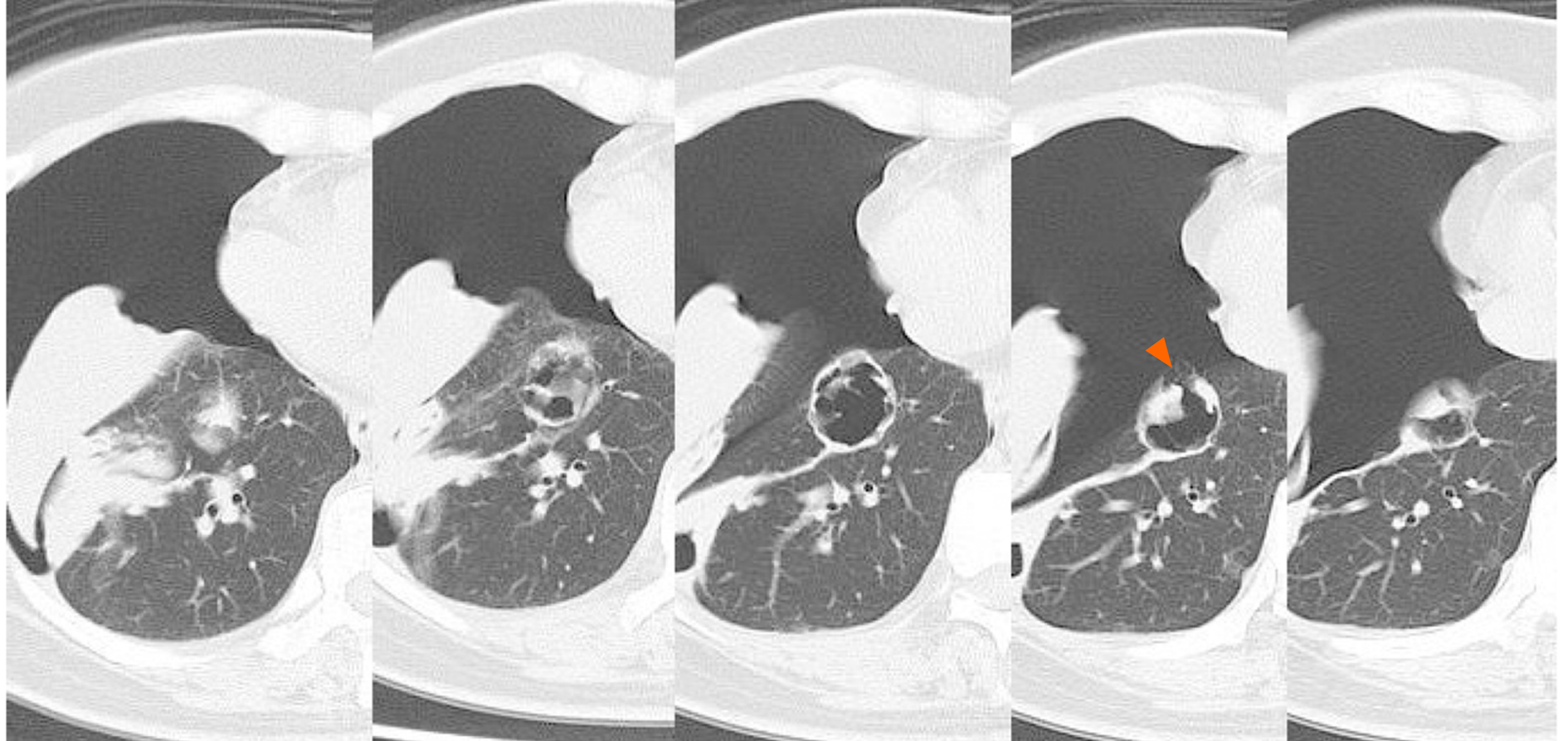
Figure 4 Chest computed tomography showing the right pneumothorax.
The cavity wall ruptured into the pleura (arrow).
- Citation: Ozaki Y, Yoshimura A, Sawaki M, Hattori M, Gondo N, Kotani H, Adachi Y, Kataoka A, Sugino K, Horisawa N, Endo Y, Nozawa K, Sakamoto S, Iwata H. Mechanisms and anatomical risk factors of pneumothorax after Bevacizumab use: A case report. World J Clin Oncol 2020; 11(7): 504-509
- URL: https://www.wjgnet.com/2218-4333/full/v11/i7/504.htm
- DOI: https://dx.doi.org/10.5306/wjco.v11.i7.504


