Copyright
©The Author(s) 2020.
World J Clin Oncol. Mar 24, 2020; 11(3): 162-168
Published online Mar 24, 2020. doi: 10.5306/wjco.v11.i3.162
Published online Mar 24, 2020. doi: 10.5306/wjco.v11.i3.162
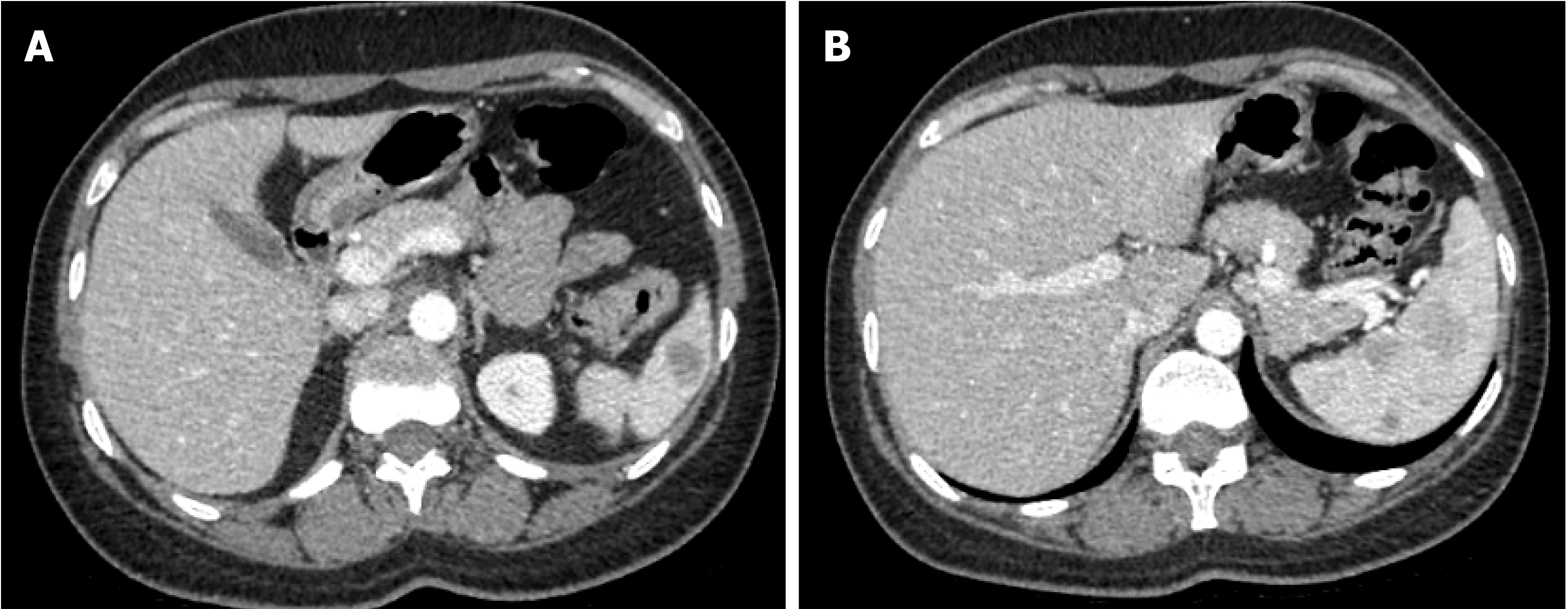
Figure 2 Computed tomography.
A, B: Spleen shows multiple circumscribed intermediate density lesions of variable size and varying enhancement, with the largest at the hilum measuring 2.4 cm × 2.1 cm and 2.7 cm × 2.1 cm. The differential diagnosis includes lymphoma, splenic peliosis, infection with multiple abscesses, and metastasis.
- Citation: Huang K, Columbie AF, Allan RW, Misra S. Thrombocytopenia with multiple splenic lesions - histiocytic sarcoma of the spleen without splenomegaly: A case report. World J Clin Oncol 2020; 11(3): 162-168
- URL: https://www.wjgnet.com/2218-4333/full/v11/i3/162.htm
- DOI: https://dx.doi.org/10.5306/wjco.v11.i3.162


