Copyright
©The Author(s) 2020.
World J Clin Oncol. Mar 24, 2020; 11(3): 162-168
Published online Mar 24, 2020. doi: 10.5306/wjco.v11.i3.162
Published online Mar 24, 2020. doi: 10.5306/wjco.v11.i3.162
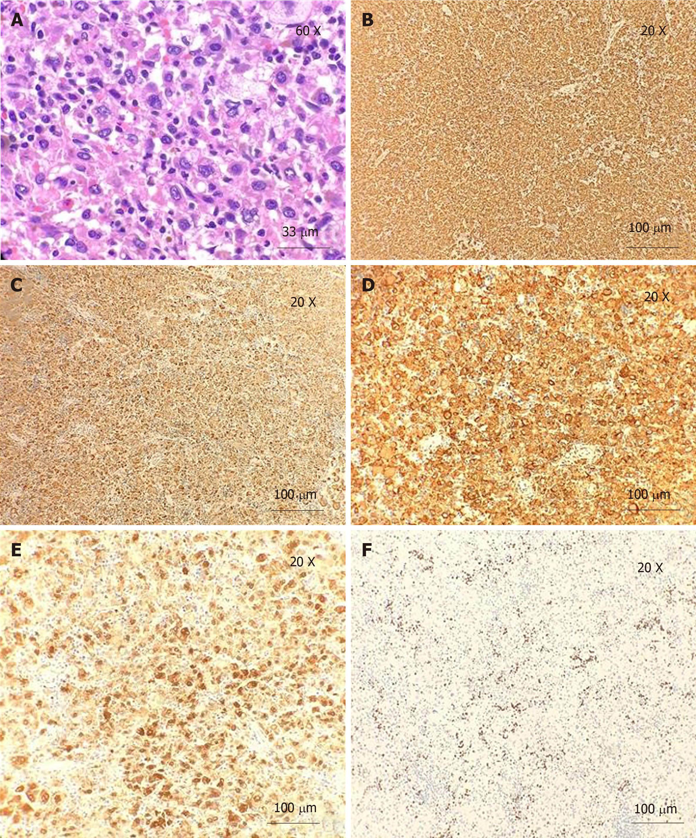
Figure 1 H and E staining and immunohistochemistry.
A: H and E staining. Neoplastic cells have abundant eosinophilic and vacuolated cytoplasm, with a large eccentric nucleus and prominent nucleoli (60 × magnification); B: Immunoreactivity for CD163 (20 × magnification); C: Immunoreactivity for CD68 (20 × magnification); D: Immunoreactivity for CD4 (20 × magnification); E: Immunoreactivity for Factor XIII (20 × magnification); F: Ki-67. Proliferation index is approximately 10% (20 × magnification).
- Citation: Huang K, Columbie AF, Allan RW, Misra S. Thrombocytopenia with multiple splenic lesions - histiocytic sarcoma of the spleen without splenomegaly: A case report. World J Clin Oncol 2020; 11(3): 162-168
- URL: https://www.wjgnet.com/2218-4333/full/v11/i3/162.htm
- DOI: https://dx.doi.org/10.5306/wjco.v11.i3.162


