Copyright
©The Author(s) 2020.
World J Clin Oncol. Feb 24, 2020; 11(2): 91-102
Published online Feb 24, 2020. doi: 10.5306/wjco.v11.i2.91
Published online Feb 24, 2020. doi: 10.5306/wjco.v11.i2.91
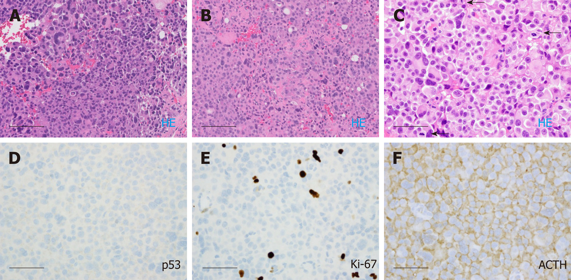
Figure 4 Histological images of the pituitary carcinoma discussed in case 2.
A: "Pituitary tumor" in 05/2010 showed an atypical pituitary adenoma with marked pleomorphism and frequent mitotic figures (ranging up to five per ten high power fields); B: The "right frontal brain lesion tissue" in 10/2016 resembled the 2010 resection material from the pituitary; C-F: The spinal cord "intradural tumor" in 11/2016 resembled the previous two resection specimens, lacked significant immunoreactivity for p53, exhibited a Ki-67 labeling index of approximately 4.8%, and showed diffuse immunoreactivity for adrenocorticotrophic hormone (arrows). Scale bar = 100 microns for panels A and B (200 ×); Scale bar = 50 microns for panels C, D, E, F (400 ×).
- Citation: Xu L, Khaddour K, Chen J, Rich KM, Perrin RJ, Campian JL. Pituitary carcinoma: Two case reports and review of literature. World J Clin Oncol 2020; 11(2): 91-102
- URL: https://www.wjgnet.com/2218-4333/full/v11/i2/91.htm
- DOI: https://dx.doi.org/10.5306/wjco.v11.i2.91


