Copyright
©The Author(s) 2020.
World J Clin Oncol. Feb 24, 2020; 11(2): 91-102
Published online Feb 24, 2020. doi: 10.5306/wjco.v11.i2.91
Published online Feb 24, 2020. doi: 10.5306/wjco.v11.i2.91
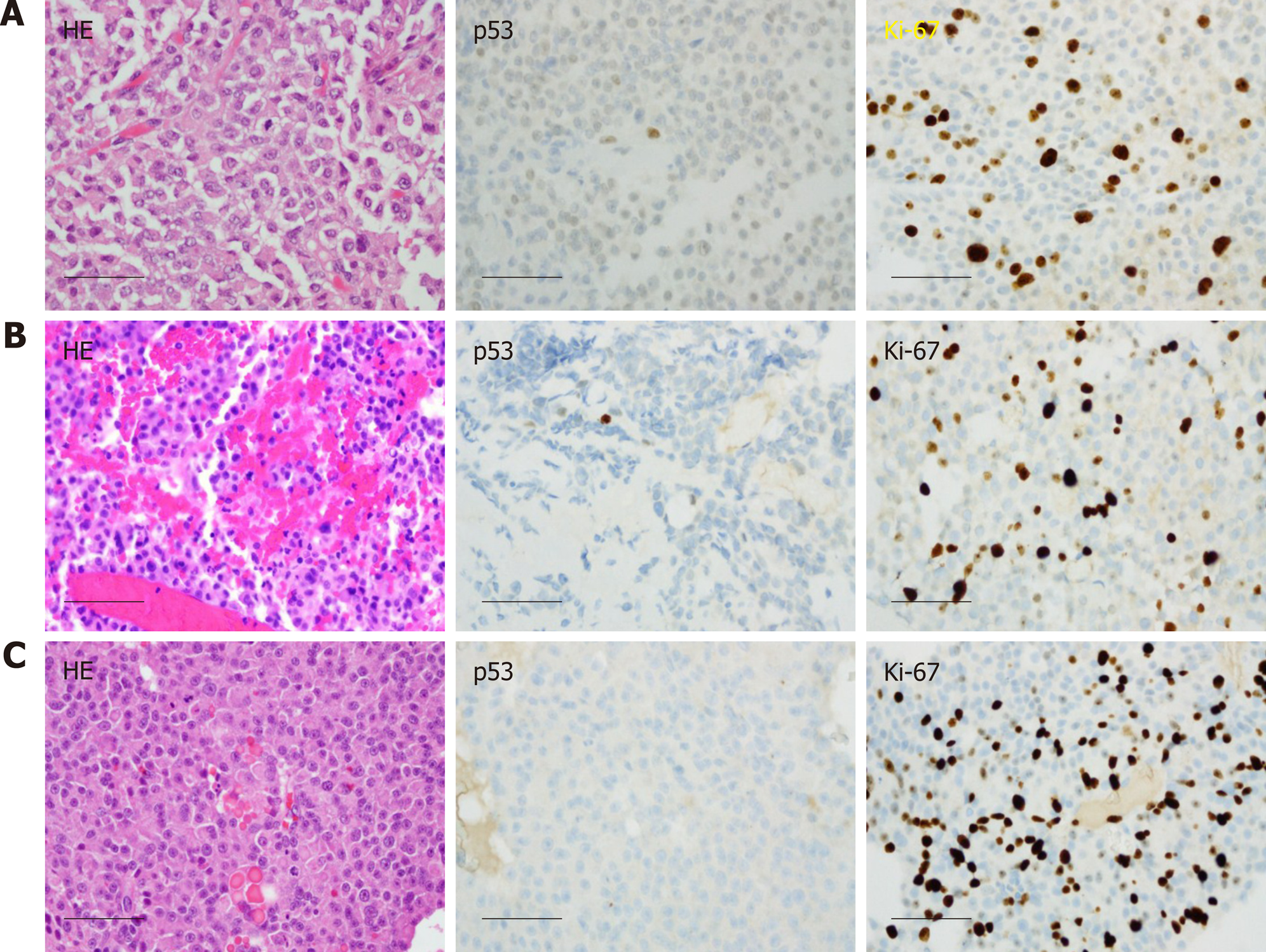
Figure 3 Histological images of the pituitary carcinoma discussed in case 1.
A: "Pituitary tumor" in 02/2011 showed an atypical pituitary adenoma with fairly frequent mitotic figures (up to four per 10 high power fields), weak immunoreactivity for p53, and a Ki-67 labeling index of 13%; B: Recurrent "pituitary tumor" in 04/2014 also showed fairly frequent mitotic figures (up to two per 10 high power fields, no significant reactivity for p53, and a Ki-67 labeling index of 17%; C: The "left convexity mass" in 05/2015 showed morphological features similar to those of the previously resected tissues, but with numerous mitotic figures (at least three per most individual high power fields), a Ki-67 labeling index of 25.5%, and no reactivity for p53. Scale bar = 50 microns for all panels (400×).
- Citation: Xu L, Khaddour K, Chen J, Rich KM, Perrin RJ, Campian JL. Pituitary carcinoma: Two case reports and review of literature. World J Clin Oncol 2020; 11(2): 91-102
- URL: https://www.wjgnet.com/2218-4333/full/v11/i2/91.htm
- DOI: https://dx.doi.org/10.5306/wjco.v11.i2.91


