Copyright
©The Author(s) 2020.
World J Clin Oncol. Feb 24, 2020; 11(2): 91-102
Published online Feb 24, 2020. doi: 10.5306/wjco.v11.i2.91
Published online Feb 24, 2020. doi: 10.5306/wjco.v11.i2.91
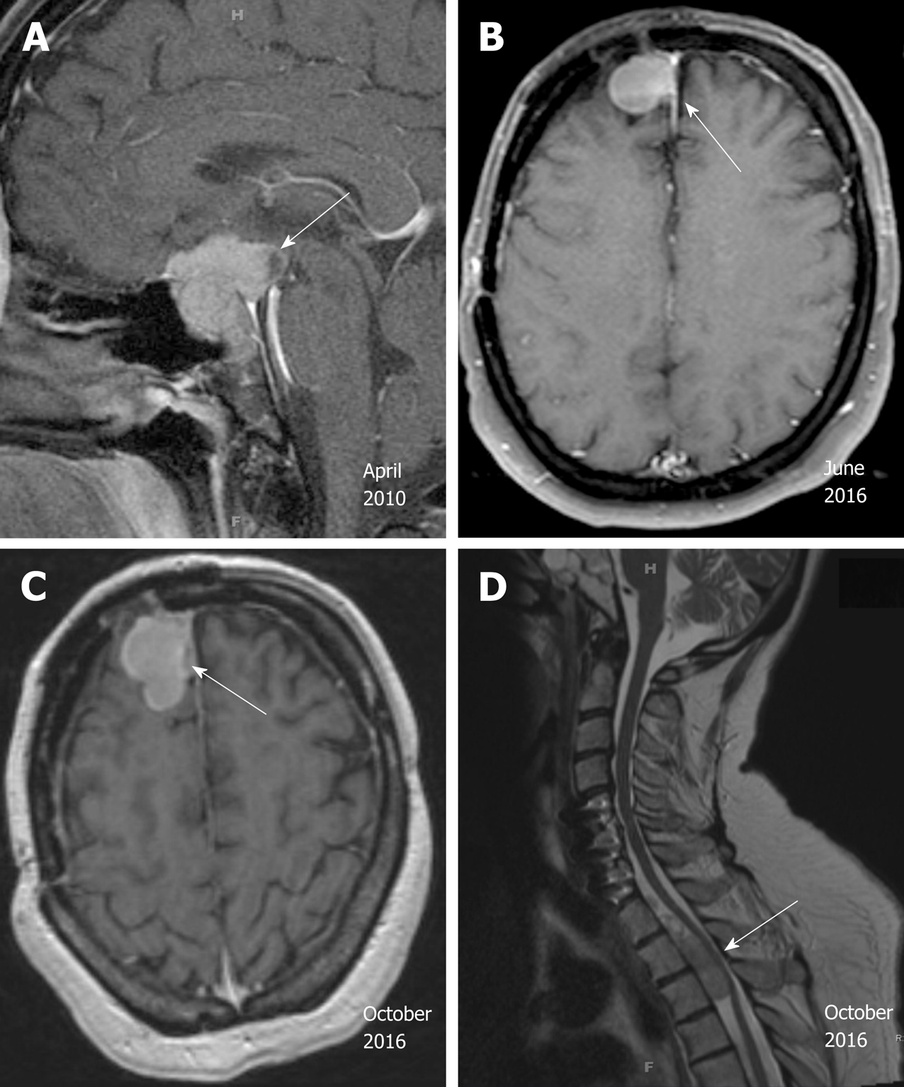
Figure 2 Radiographic images of the pituitary carcinoma discussed in case 2.
A: Magnetic resonance imaging (MRI) in April 2010 shows a suprasellar mass invading the optic chiasm and enveloping the carotid siphon and cavernous sinus (arrow); B: MRI in June 2016 shows recurrence of a 2.3 cm right frontal lesion (arrow); C: MRI of brain in October 2016 shows enhancing anterior right frontal lobe mass measuring 4.1 cm × 2.1 cm (arrow); D: MRI of spine in October 2016 shows extramedullary metastatic tumor within the ventral spinal canal at T1-T3 levels (arrow, T5 lesion is not shown here).
- Citation: Xu L, Khaddour K, Chen J, Rich KM, Perrin RJ, Campian JL. Pituitary carcinoma: Two case reports and review of literature. World J Clin Oncol 2020; 11(2): 91-102
- URL: https://www.wjgnet.com/2218-4333/full/v11/i2/91.htm
- DOI: https://dx.doi.org/10.5306/wjco.v11.i2.91


