Copyright
©The Author(s) 2020.
World J Clin Oncol. Feb 24, 2020; 11(2): 83-90
Published online Feb 24, 2020. doi: 10.5306/wjco.v11.i2.83
Published online Feb 24, 2020. doi: 10.5306/wjco.v11.i2.83
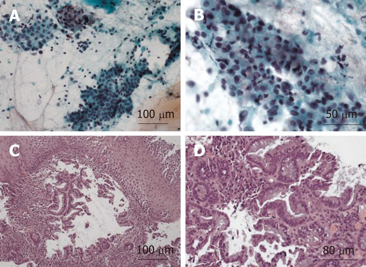
Figure 4 Histologic preparation of right cervical lymph node core needle biopsy.
A: Immunohistochemical stain for Caudal Type Homeobox 2 positive stain, 10 × magnifications; B: Immunohistochemical stain for thyroid transcription factor 1 weakly positive stain, 40 × magnification; C: Loss of lymph node architecture with several glands present on a background of fibrous stroma, 10 × magnification; D: Higher power view demonstrating cytologic atypia and abundant mucin, 20 × magnification.
- Citation: Burns EA, Kasparian S, Khan U, Abdelrahim M. Pancreatic adenocarcinoma with early esophageal metastasis: A case report and review of literature. World J Clin Oncol 2020; 11(2): 83-90
- URL: https://www.wjgnet.com/2218-4333/full/v11/i2/83.htm
- DOI: https://dx.doi.org/10.5306/wjco.v11.i2.83


