Copyright
©The Author(s) 2020.
World J Clin Oncol. Feb 24, 2020; 11(2): 83-90
Published online Feb 24, 2020. doi: 10.5306/wjco.v11.i2.83
Published online Feb 24, 2020. doi: 10.5306/wjco.v11.i2.83
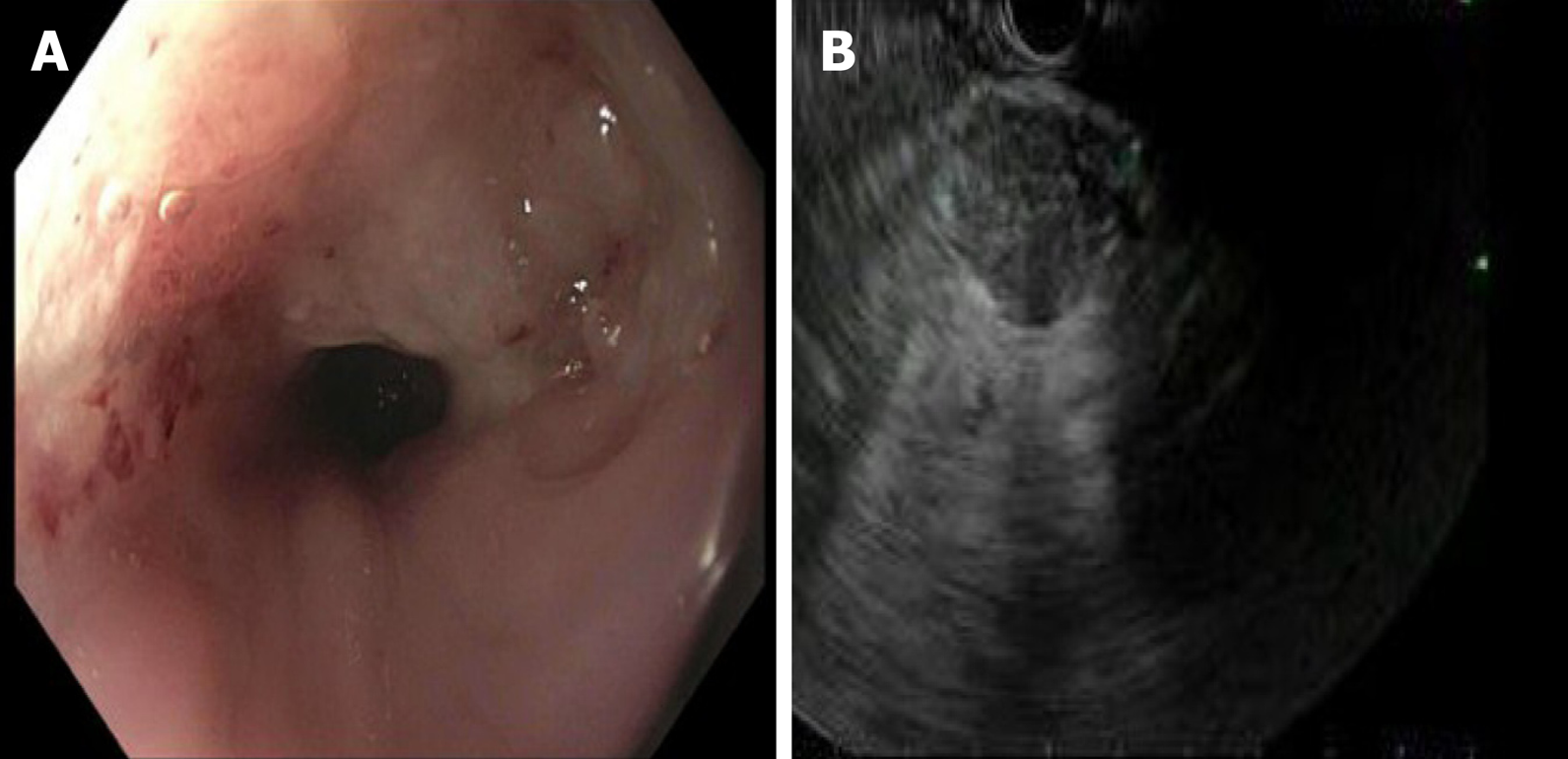
Figure 3 Upper endoscopy which demonstrated a mass in the upper third of the esophagus.
A: Upper endoscopy with a non-obstructing non-circumferential submucosal mass in the upper third of the esophagus; B: Endoscopy ultrasonography demonstrating 2.5 cm × 2.4 cm heterogenous, hypoechoic solid mass with irregular outer borders in the body of the pancreas.
- Citation: Burns EA, Kasparian S, Khan U, Abdelrahim M. Pancreatic adenocarcinoma with early esophageal metastasis: A case report and review of literature. World J Clin Oncol 2020; 11(2): 83-90
- URL: https://www.wjgnet.com/2218-4333/full/v11/i2/83.htm
- DOI: https://dx.doi.org/10.5306/wjco.v11.i2.83


