Copyright
©The Author(s) 2020.
World J Clin Oncol. Feb 24, 2020; 11(2): 83-90
Published online Feb 24, 2020. doi: 10.5306/wjco.v11.i2.83
Published online Feb 24, 2020. doi: 10.5306/wjco.v11.i2.83
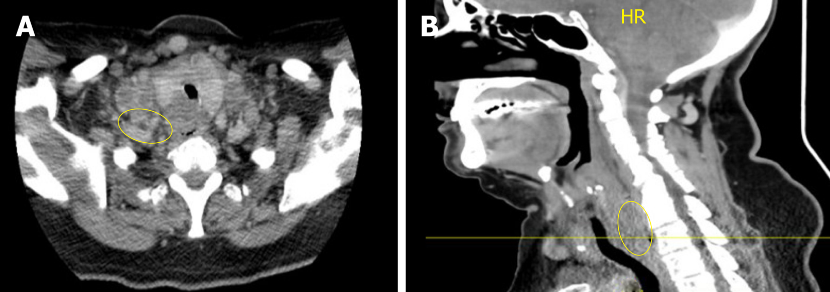
Figure 1 Computed tomography of the neck and chest.
A: Axial view; B: Sagittal View. Heterogeneous and partially necrotic mass like enhancement dorsal to the larynx and upper trachea which appears to dorsally displace the visible portions of the esophagus. The mass measures approximately 3 cm transverse by 1.8 cm anterior-posteriorly, and extends craniocaudally into the superior mediastinum. There is Widespread metastatic and necrotic adenopathy involving cervical lymph nodes.
- Citation: Burns EA, Kasparian S, Khan U, Abdelrahim M. Pancreatic adenocarcinoma with early esophageal metastasis: A case report and review of literature. World J Clin Oncol 2020; 11(2): 83-90
- URL: https://www.wjgnet.com/2218-4333/full/v11/i2/83.htm
- DOI: https://dx.doi.org/10.5306/wjco.v11.i2.83


