Copyright
©The Author(s) 2020.
World J Clin Oncol. Feb 24, 2020; 11(2): 103-109
Published online Feb 24, 2020. doi: 10.5306/wjco.v11.i2.103
Published online Feb 24, 2020. doi: 10.5306/wjco.v11.i2.103
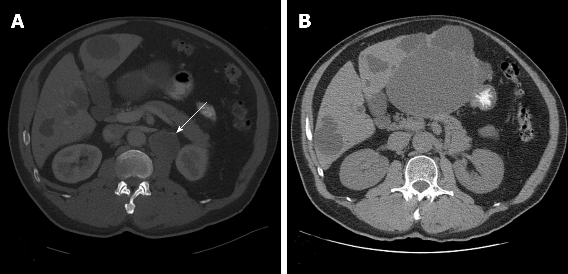
Figure 1 A computed tomography scan of the abdomen and pelvis.
A: A 1.8 cm left retroperitoneal lymph node which was concerning for malignancy (arrow); B: No recurrent or metastatic disease and no evidence of retroperitoneal adenopathy observed 5 years following the lymph node dissection.
- Citation: Shields LB, Kalebasty AR. Metastatic clear cell renal cell carcinoma in isolated retroperitoneal lymph node without evidence of primary tumor in kidneys: A case report. World J Clin Oncol 2020; 11(2): 103-109
- URL: https://www.wjgnet.com/2218-4333/full/v11/i2/103.htm
- DOI: https://dx.doi.org/10.5306/wjco.v11.i2.103


