Copyright
©The Author(s) 2019.
World J Clin Oncol. Jul 24, 2019; 10(7): 269-278
Published online Jul 24, 2019. doi: 10.5306/wjco.v10.i7.269
Published online Jul 24, 2019. doi: 10.5306/wjco.v10.i7.269
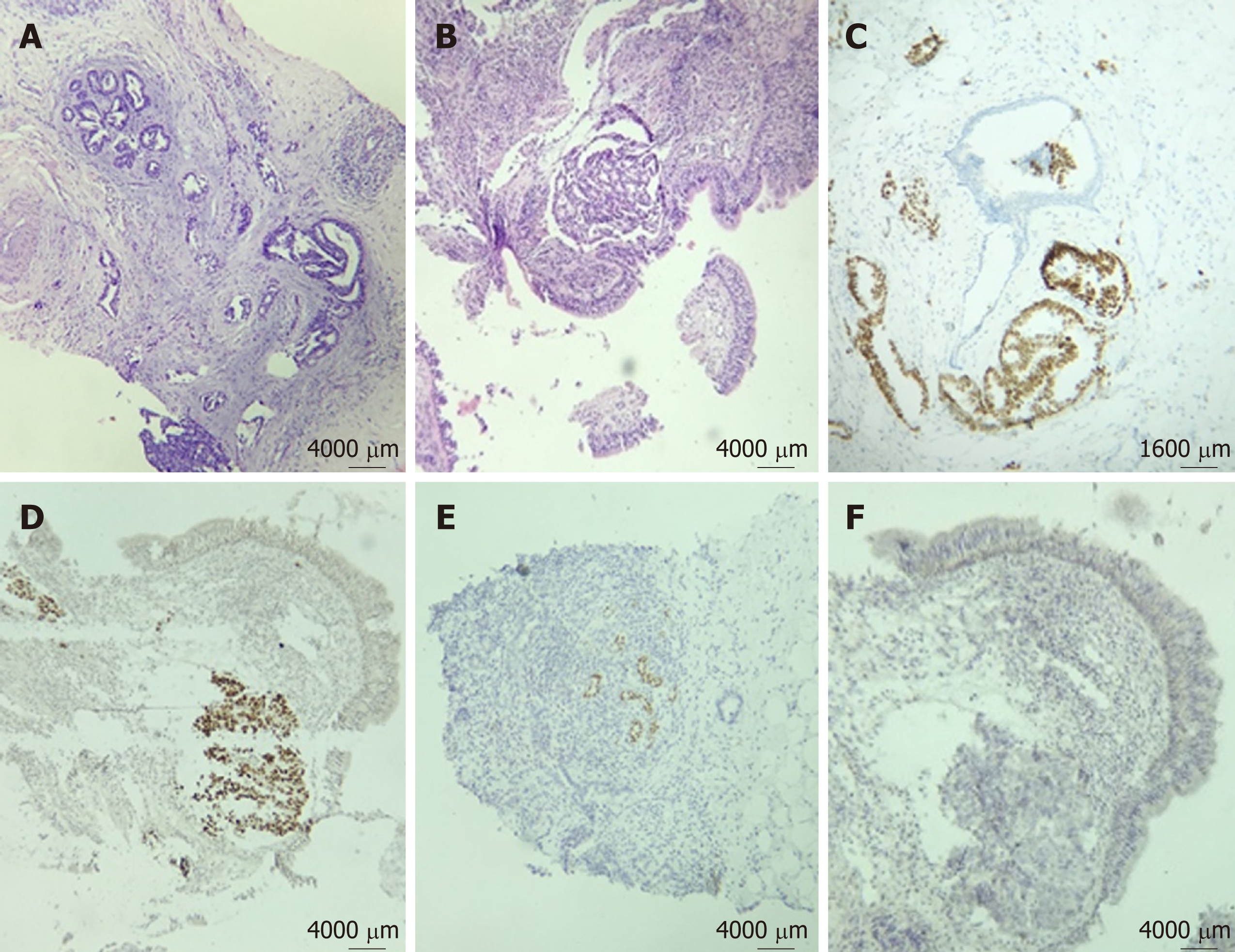
Figure 3 Breast biopsy showed adenocarcinoma infiltrating into the adjacent parenchyma.
A: Ducts were not involved by the tumor, and no evidence of in situ carcinoma was obtained (x 40); B: Bronchoscopy biopsy (HE) showed poorly differentiated adenocarcinoma (x 40); C, D: Immunohistochemical staining for thyroid transcription factor-1 was positive on both breast (C) and lung specimens (D); E, F: GATA3 staining was negative in both breast (E) and lung tissue (F).
- Citation: Enrico D, Saucedo S, Bravo I. Breast metastasis from primary lung adenocarcinoma in a young woman: A case report and literature review. World J Clin Oncol 2019; 10(7): 269-278
- URL: https://www.wjgnet.com/2218-4333/full/v10/i7/269.htm
- DOI: https://dx.doi.org/10.5306/wjco.v10.i7.269


