Copyright
©2010 Baishideng Publishing Group Co.
World J Clin Oncol. Nov 10, 2010; 1(1): 3-11
Published online Nov 10, 2010. doi: 10.5306/wjco.v1.i1.3
Published online Nov 10, 2010. doi: 10.5306/wjco.v1.i1.3
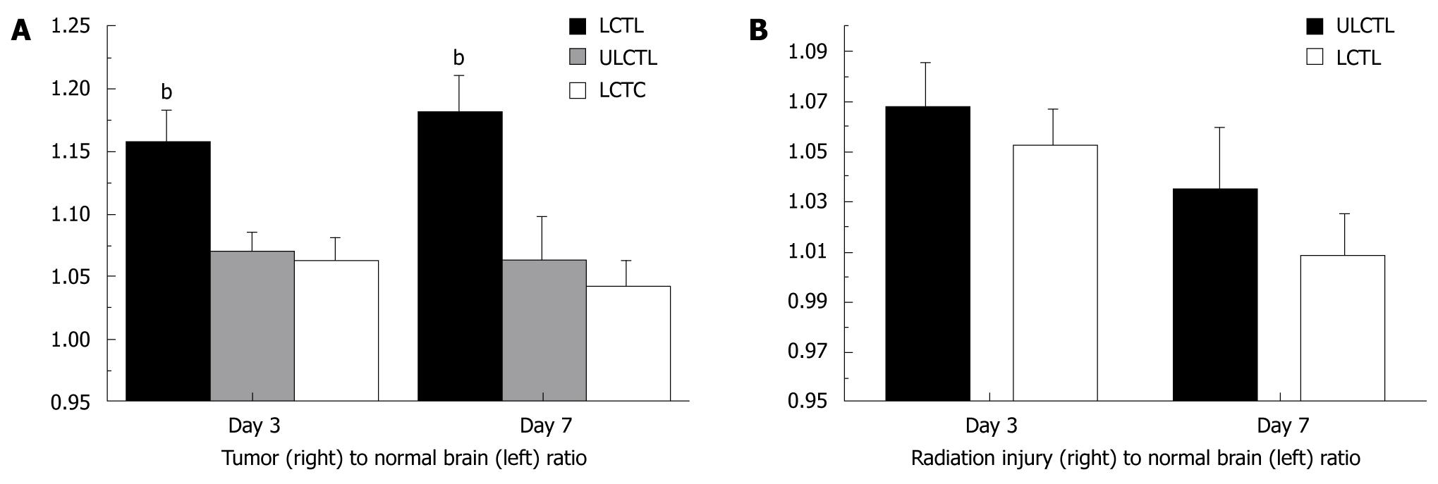
Figure 4 Accumulation of labeled cytotoxic T-cells in implanted U251 tumor and radiation injured sites in rat brain.
A: Analyses of R2* values normalized to contralateral normal hemisphere (indirect indicator of the accumulation of iron positive cells) showed significantly higher (bP≤ 0.001) accumulation of iron positive cells in tumor that received labeled cytotoxic T-cells (LCTL) compared to that of labeled control T-cells (LCTC) and unlabeled CTLs (ULCTL). The number of accumulated cells was higher at both day 3 and 7; B: Similar analyses of R2* values in radiation injured brain normalized to contralateral normal hemisphere showed no difference between the groups of animals that received labeled and unlabeled CTLs.
- Citation: Arbab AS. Cytotoxic T-cells as imaging probes for detecting glioma. World J Clin Oncol 2010; 1(1): 3-11
- URL: https://www.wjgnet.com/2218-4333/full/v1/i1/3.htm
- DOI: https://dx.doi.org/10.5306/wjco.v1.i1.3


