Copyright
©The Author(s) 2017.
World J Gastrointest Pathophysiol. Aug 15, 2017; 8(3): 142-149
Published online Aug 15, 2017. doi: 10.4291/wjgp.v8.i3.142
Published online Aug 15, 2017. doi: 10.4291/wjgp.v8.i3.142
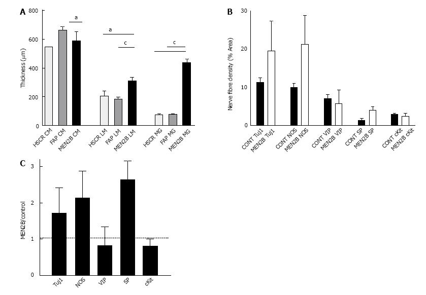
Figure 2 Thickness of muscle layers and ganglia and density of nerve fibres in normal colon and MEN2B colon.
A: Comparison of thickness (mm) of muscle and myenteric ganglia in colon from Control (HSCR and FAP) and MEN2B patient. Graph shows mean and SEM. HSCR n = 6, FAP n = 12, MEN2B n = 24, aP < 0.05, cP < 0.001; B: Nerve fibre and ICC density in circular muscle in transverse colon from Control (HSCR and FAP combined, n = 3) and MEN2B patient (n = 3). Percent area of circular muscle containing immunoreactive pixels; C: Relative nerve fibre and ICC density in the circular muscle in MEN2B patient (n = 3) relative to Control patients (HSCR and FAP combined, n = 3). Labelling with Tuj1, NOS, VIP, SP and cKit (for interstitial cells of Cajal).
- Citation: Hutson JM, Farmer PJ, Peck CJ, Chow CW, Southwell BR. Multiple endocrine neoplasia 2B: Differential increase in enteric nerve subgroups in muscle and mucosa. World J Gastrointest Pathophysiol 2017; 8(3): 142-149
- URL: https://www.wjgnet.com/2150-5330/full/v8/i3/142.htm
- DOI: https://dx.doi.org/10.4291/wjgp.v8.i3.142


