Copyright
©The Author(s) 2017.
World J Gastrointest Pathophysiol. Aug 15, 2017; 8(3): 142-149
Published online Aug 15, 2017. doi: 10.4291/wjgp.v8.i3.142
Published online Aug 15, 2017. doi: 10.4291/wjgp.v8.i3.142
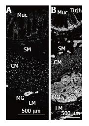
Figure 1 Nerve fibres in normal colon and MEN2B colon.
Tuj1 immunoreactivity showing nerve fibres in full thickness biopsy of the transverse colon cut with circular muscle in cross section. A: Control patient (4-year-old, HSCR normal margin) showing multiple nerve fibres in the mucosa (Muc) within and at the base of the crypts, small ganglia in the submucosa (SM), nerve fibres in cross section in the circular muscle (CM) and myenteric ganglia (MG) between the CM and longitudinal muscle layer (LM); B: MEN2B patient showing greatly increased amount of labelling and increase in size of MG and SM ganglia. The MG is increased in diameter to a thickness similar to the circular muscle. The density and number of nerve fibres in the CM is higher than control. The nerve fibres in the Muc are greatly increased. The SM contains many large ganglia near the CM, near the Muc and in between. Neurons are not distinguishable because this antibody only displays nerve fibres. Note A and B are at the same magnification. CM: Circular mucle; LM: Longitudinal muscle; MG: Myenteric ganglia.
- Citation: Hutson JM, Farmer PJ, Peck CJ, Chow CW, Southwell BR. Multiple endocrine neoplasia 2B: Differential increase in enteric nerve subgroups in muscle and mucosa. World J Gastrointest Pathophysiol 2017; 8(3): 142-149
- URL: https://www.wjgnet.com/2150-5330/full/v8/i3/142.htm
- DOI: https://dx.doi.org/10.4291/wjgp.v8.i3.142


