Copyright
©The Author(s) 2017.
World J Gastrointest Pathophysiol. May 15, 2017; 8(2): 39-50
Published online May 15, 2017. doi: 10.4291/wjgp.v8.i2.39
Published online May 15, 2017. doi: 10.4291/wjgp.v8.i2.39
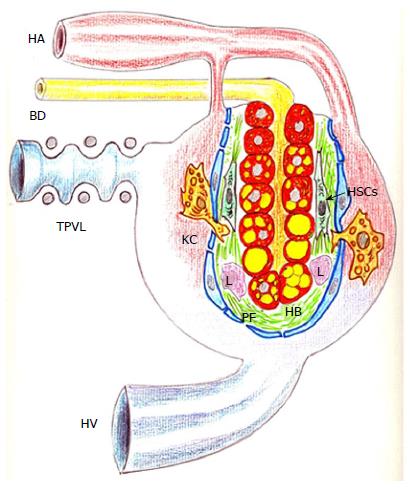
Figure 3 Schematic representation of the liver parenchyma after partial portal vein in the rat.
The deposit of lipids within the hepatocytes and the liver arterialization, associated with defenestration of the sinusoidal endothelium, and the perisinuosidal fibrosis stand out. BD: Bile duct; HA: Hepatic artery; HV: Hepatic vein; HB: Hepatocyte ballooning; HSCs: Hepatic stellate cells; KC: Kupffer cell; L: Leukocyte; PF: Perisinuosidal fibrosis; TPVL: Triple partial portal vein ligation.
- Citation: Aller MA, Arias N, Peral I, García-Higarza S, Arias JL, Arias J. Embrionary way to create a fatty liver in portal hypertension. World J Gastrointest Pathophysiol 2017; 8(2): 39-50
- URL: https://www.wjgnet.com/2150-5330/full/v8/i2/39.htm
- DOI: https://dx.doi.org/10.4291/wjgp.v8.i2.39


