Copyright
©The Author(s) 2017.
World J Gastrointest Pathophysiol. May 15, 2017; 8(2): 39-50
Published online May 15, 2017. doi: 10.4291/wjgp.v8.i2.39
Published online May 15, 2017. doi: 10.4291/wjgp.v8.i2.39
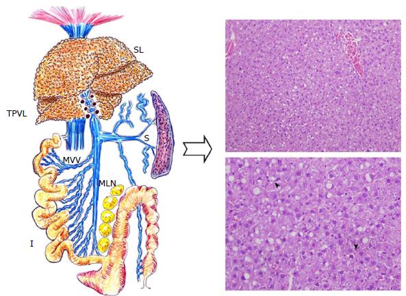
Figure 1 Liver histopathological changes of a rat after three months with triple partial portal vein ligation.
Hepatic steatotic areas are mainly distributed in zone 1 of the liver acinus, but tipically hepatocyte ballooning is more apparent near zone 3. Both macrovesicular and microvesicular steatosis are evident as well as scattered necroinflammatory foci (arrowheads) (H and E stain, × 200). I: Intestine; MVV: Mesenteric venous vasculopathy; S: Spleen; SL: Steatotic liver; TPVL: Triple partial portal vein ligation.
- Citation: Aller MA, Arias N, Peral I, García-Higarza S, Arias JL, Arias J. Embrionary way to create a fatty liver in portal hypertension. World J Gastrointest Pathophysiol 2017; 8(2): 39-50
- URL: https://www.wjgnet.com/2150-5330/full/v8/i2/39.htm
- DOI: https://dx.doi.org/10.4291/wjgp.v8.i2.39


