Copyright
©The Author(s) 2016.
World J Gastrointest Pathophysiol. Feb 15, 2016; 7(1): 138-149
Published online Feb 15, 2016. doi: 10.4291/wjgp.v7.i1.138
Published online Feb 15, 2016. doi: 10.4291/wjgp.v7.i1.138
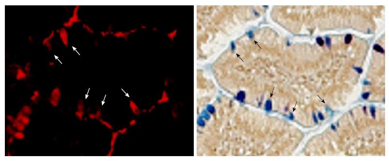
Figure 3 Amylase is expressed in a subset of goblet cells.
Immunofluorescence of amylase (top, red) identifies many, but not all, goblet cells that are clearly stained with AB/PAS (bottom). The same sections (slides) from NKCC1-null intestine was used for immunofluorescence, imaged, and then stained with AB/PAS. Arrows indicate the same cell in both images. AB/PAS: Alcian Blue/Periodic Acid-Schiff.
- Citation: Bradford EM, Vairamani K, Shull GE. Differential expression of pancreatic protein and chemosensing receptor mRNAs in NKCC1-null intestine. World J Gastrointest Pathophysiol 2016; 7(1): 138-149
- URL: https://www.wjgnet.com/2150-5330/full/v7/i1/138.htm
- DOI: https://dx.doi.org/10.4291/wjgp.v7.i1.138


