Copyright
©The Author(s) 2016.
World J Gastrointest Pathophysiol. Feb 15, 2016; 7(1): 138-149
Published online Feb 15, 2016. doi: 10.4291/wjgp.v7.i1.138
Published online Feb 15, 2016. doi: 10.4291/wjgp.v7.i1.138
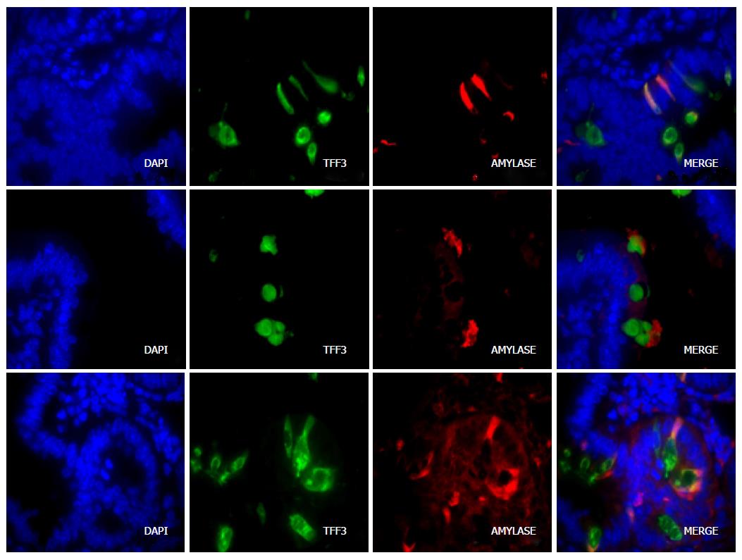
Figure 2 Colocalization of peptidases in goblet cells of the duodenum.
DAPI (blue) identifies nuclei and TFF3 (intestinal trefoil factor, green fluorescence) identifies goblet cells. Elastase, amylase and trypsin are identified by red fluorescence. The merged images show colocalization of each of these proteases and TFF3. Staining patterns were assessed in 4 wild-type and 3 Nkcc1-/- mice. The images shown are for NKCC1-null mice. Control sections with only primary or only secondary antibodies do not identify goblet cells (not shown).
- Citation: Bradford EM, Vairamani K, Shull GE. Differential expression of pancreatic protein and chemosensing receptor mRNAs in NKCC1-null intestine. World J Gastrointest Pathophysiol 2016; 7(1): 138-149
- URL: https://www.wjgnet.com/2150-5330/full/v7/i1/138.htm
- DOI: https://dx.doi.org/10.4291/wjgp.v7.i1.138


