Copyright
©2014 Baishideng Publishing Group Inc.
World J Gastrointest Pathophysiol. Aug 15, 2014; 5(3): 252-270
Published online Aug 15, 2014. doi: 10.4291/wjgp.v5.i3.252
Published online Aug 15, 2014. doi: 10.4291/wjgp.v5.i3.252
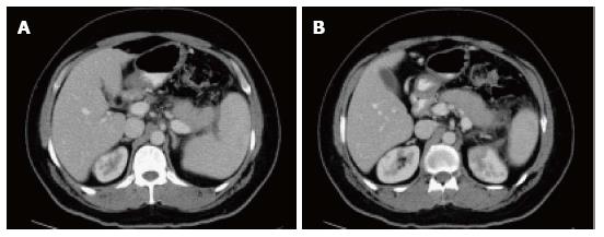
Figure 19 Autoimmune pancreatitis.
A, B: Axial CT scan during the late arterial phase. There is evidence of diffuse pancreatic swelling with loss of the normal pancreatic lobulation, obliteration of the pancreatic duct and subtle low attenuating peripancreatic rim (A, B) in keeping with autoimmune pancreatitis. Patient had high IgG4 level (> 0.500 g/L).
- Citation: Busireddy KK, AlObaidy M, Ramalho M, Kalubowila J, Baodong L, Santagostino I, Semelka RC. Pancreatitis-imaging approach. World J Gastrointest Pathophysiol 2014; 5(3): 252-270
- URL: https://www.wjgnet.com/2150-5330/full/v5/i3/252.htm
- DOI: https://dx.doi.org/10.4291/wjgp.v5.i3.252


