Copyright
©2014 Baishideng Publishing Group Inc.
World J Gastrointest Pathophysiol. Aug 15, 2014; 5(3): 252-270
Published online Aug 15, 2014. doi: 10.4291/wjgp.v5.i3.252
Published online Aug 15, 2014. doi: 10.4291/wjgp.v5.i3.252
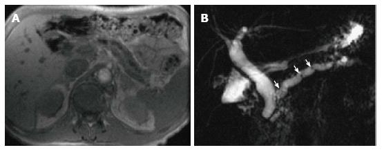
Figure 18 Chronic pancreatitis.
A: Axial T1-weighted GRE MRI. B: Coronal-oblique thick-slab MRCP image. There is evidence of diffuse thinning of the pancreatic parenchyma with uniform dilatation of the pancreatic duct and prominence of the pancreatic duct side-branches (A-B), associated with multiple tiny stones at the proximal pancreatic duct (arrows, B) in keeping with chronic pancreatitis. There is also mild uniform dilatation of the CBD, which tapers down to the level of the pancreatic duct (B).
- Citation: Busireddy KK, AlObaidy M, Ramalho M, Kalubowila J, Baodong L, Santagostino I, Semelka RC. Pancreatitis-imaging approach. World J Gastrointest Pathophysiol 2014; 5(3): 252-270
- URL: https://www.wjgnet.com/2150-5330/full/v5/i3/252.htm
- DOI: https://dx.doi.org/10.4291/wjgp.v5.i3.252


