Copyright
©2014 Baishideng Publishing Group Inc.
World J Gastrointest Pathophysiol. Aug 15, 2014; 5(3): 252-270
Published online Aug 15, 2014. doi: 10.4291/wjgp.v5.i3.252
Published online Aug 15, 2014. doi: 10.4291/wjgp.v5.i3.252
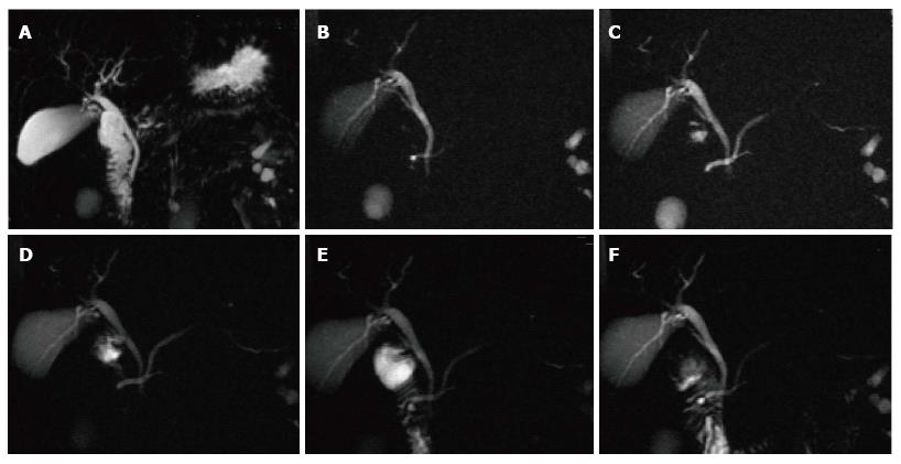
Figure 16 Pancreatic divisum, with a small Santorinicele.
A: Coronal 3D- maximum intensity projection MRCP image before administration of secretin; B-F: Selected dynamic secretin thick-slab MRCP images obtained at 30 s (B), 60 s (C), 120 s (D), 4 min (E) and 9 min (F). Prior to administration of secretin, it is difficult to identify the main pancreatic duct (A). After administration of secretin, there is better delineation of the main pancreatic duct (C), with demonstration of pancreatic divisum. There is also enlargement of the accessory pancreatic duct, with demonstration of a small santorinicele (B, F). S-MRCP allows qualitative and quantitative assessment of pancreatic exocrine secretions. In this case, the pancreatic flow output was considered within normal limits; excluding early chronic pancreatitis.
- Citation: Busireddy KK, AlObaidy M, Ramalho M, Kalubowila J, Baodong L, Santagostino I, Semelka RC. Pancreatitis-imaging approach. World J Gastrointest Pathophysiol 2014; 5(3): 252-270
- URL: https://www.wjgnet.com/2150-5330/full/v5/i3/252.htm
- DOI: https://dx.doi.org/10.4291/wjgp.v5.i3.252


