Copyright
©2014 Baishideng Publishing Group Inc.
World J Gastrointest Pathophysiol. Aug 15, 2014; 5(3): 252-270
Published online Aug 15, 2014. doi: 10.4291/wjgp.v5.i3.252
Published online Aug 15, 2014. doi: 10.4291/wjgp.v5.i3.252
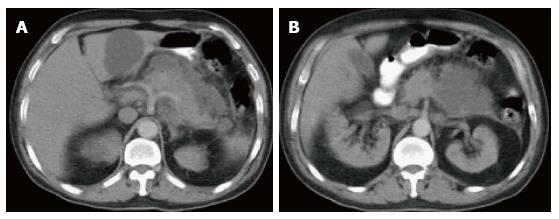
Figure 9 Acute interstitial edematous pancreatitis and acute peripancreatic fluid collections.
A-B: Axial CT scan during the portal venous phase. The pancreas is mildly thickened and demonstrates mildly heterogenous enhancement, reflective of edema, in keeping with acute interstitial edematous pancreatitis. There is a peripancreatic fluid with imperceptible wall in keeping with acute peripancreatic fluid collections.
- Citation: Busireddy KK, AlObaidy M, Ramalho M, Kalubowila J, Baodong L, Santagostino I, Semelka RC. Pancreatitis-imaging approach. World J Gastrointest Pathophysiol 2014; 5(3): 252-270
- URL: https://www.wjgnet.com/2150-5330/full/v5/i3/252.htm
- DOI: https://dx.doi.org/10.4291/wjgp.v5.i3.252


